Published online Jul 7, 2006. doi: 10.3748/wjg.v12.i25.4096
Revised: March 15, 2006
Accepted: March 27, 2006
Published online: July 7, 2006
Metastatic tumors of the gastrointestinal tract are rare. We describe a case of gastric metastasis due to primary lung cancer, revealed by an upper gastrointestinal endoscopy (UGIE). Haematogenous metastases to the stomach are a rare event. To our knowledge, only 55 cases have been described in the international literature. In these patients, the prognosis is very poor. We report herein a case of gastric metastasis by lung small cell carcinoma, with a review of the literature about this rare entity.
- Citation: Casella G, Bella CD, Cambareri AR, Buda CA, Corti G, Magri F, Crippa S, Baldini V. Gastric metastasis by lung small cell carcinoma. World J Gastroenterol 2006; 12(25): 4096-4097
- URL: https://www.wjgnet.com/1007-9327/full/v12/i25/4096.htm
- DOI: https://dx.doi.org/10.3748/wjg.v12.i25.4096
Metastatic tumors of the gastrointestinal tract are rare[1]. In most cases, the diagnosis is possible only at autopsy. To our best of knowledge, only 55 cases have been described in international literature so far[2].
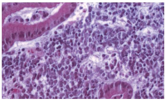
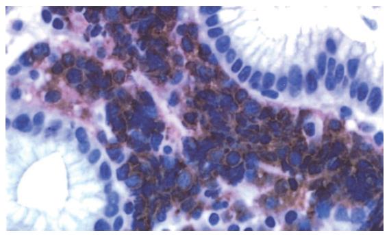
We describe a case of gastric metastasis due to primary lung cancer revealed by an upper gastrointestinal endoscopy (UGIE). In March 2005, a 63-year-old Caucasian male was referred to our department because of fever (38°C), weight loss (5 kg in few months), epigastric pain, and constipation. He had an accidental chest trauma associated to right rib fractures 3 years back. He was a smoker of 20 cigarettes per day. At admission to our department, his general conditions were poor. Lymphadenopathy (> 3 cm) in the left scalenic region, and liver enlargement were evident. A mild anaemia (Hb 110 g/L), low platelet count (40 × 109/L), and a marked increase of lactic dehydrogenase (LDH > 1000 U/L) were the most important blood biochemical alterations. A chest x-ray revealed a lung mass (> 5 cm in diameter) in the upper left hilar region. Multiple 1-3 cm secondary liver lesions scattered in both lobes, and with a typical “bull’s eye” appearance were revealed by ultrasonography. An UGIE showed a 1.5-cm raised area, depressed on the tip with recent signs of haemorrhage in the gastric corpus, which was histologically identified as the metastasis of an undifferentiated small cell carcinoma (Figure 1). A total body computed tomography (CT) confirmed a left lung neoplasm associated to an infiltration of the left pulmonary artery, left upper bronchus stenosis, bulk mediastinal lymphadenopathies (> 3 cm), and bilateral pleural effusions. Liver secondaries were confirmed and multiple brain metastases were localised in the left parietal lobe and, bilaterally, in the occipital lobes. A total body bone scan showed diffuse bone involvement. A bronchoscopy and percutaneous biopsy of the left supraclavicular lymph node confirmed the diagnosis of diffuse poorly differentiated primary lung small cell cancer (Figure 2). The patient was provided only with a supportive care, considering his poor condition. He died one month later.
Haematogenous metastases to the stomach is a rare event[3]. The most frequent tumors involved in secondary gastric sites are melanoma, breast, and lung cancer. Most patients with gastrointestinal metastases are asymptomatic[1]. At autopsy, gastric metastases are present in 14% of all patients with lung cancer[4]. Lung small cell carcinoma has a high incidence of vascular invasion[3] and the gastric submucosal layer is always involved, followed by the mucosa, muscularis propria, and serosa[5]. Complete inclusion of the metastasis within the gastric wall may be a sign of a primary tumor of another origin[5].
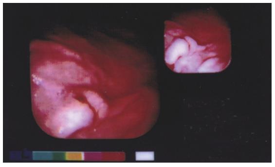
Abdominal pain is the most frequent (80% of the cases) symptom in the symptomatic patient. Kadakia et al[1] also reported iron-deficiency anaemia and acute upper GI bleeding. Severe upper gastrointestinal bleeding, hypovolemic shock, and stomach perforation may be the cause of death of the patient. Differential diagnosis with lymphoma, ectopic pancreas, carcinoid tumor, eosinophilic granuloma, Kaposi’s sarcoma and gastric ulcer is important[3]. Gastric metastatic lesions have a particular endoscopic aspect, appearing as ”volcano-like” or umbilicated on the tip[3] (Figure 3). These lesions may present with three different appearances[3]: (1) multiple nodules of variable size with a central ulcer; (2) submucosal, raised, and ulcerated at the tip and defined as “volcano-like”; and (3) raised areas without a central ulcer. In few cases, the lesions appear as polyps or raised plaques[3]. It has been reported a high percentage of gastric metastases in the fundus and cardias[4], but in our experience, the lesions are present in the gastric corpus region, which is in agreement with Kadakia et al[1]. Hamatake et al[6] described a rare case of solitary gastric metastasis from the lower left lung. This was a poorly differentiated adenocarcinoma diagnosed during UGIE for UGI bleeding, which resolved after gastrectomy and left lobectomy.
It is important to identify these lesions in order to prevent serious complications, such as gastrointestinal bleeding, viscus perforation, or gastric outlet obstruction[4]. An upper gastrointestinal endoscopy should be recommended before starting chemotherapy[5], as confirmed by Suzaki et al[7] who described a case of gastric perforation during systemic chemotherapy for lung neoplasm due to metastasis[7] .
S- Editor Wang J L- Editor Kumar M E- Editor Ma WH
| 1. | Kadakia SC, Parker A, Canales L. Metastatic tumors to the upper gastrointestinal tract: endoscopic experience. Am J Gastroenterol. 1992;87:1418-1423. [PubMed] |
| 2. | Oh JC, Lee GS, Kim JS, Park Y, Lee SH, Kim A, Lee JM, Kim KS. [A case of gastric metastasis from small cell lung carcinoma]. Korean J Gastroenterol. 2004;44:168-171. [PubMed] |
| 3. | Green LK. Hematogenous metastases to the stomach. A review of 67 cases. Cancer. 1990;65:1596-1600. [RCA] [PubMed] [DOI] [Full Text] [Cited by in RCA: 2] [Reference Citation Analysis (0)] |
| 4. | Antler AS, Ough Y, Pitchumoni CS, Davidian M, Thelmo W. Gastrointestinal metastases from malignant tumors of the lung. Cancer. 1982;49:170-172. [RCA] [PubMed] [DOI] [Full Text] [Cited by in RCA: 1] [Reference Citation Analysis (0)] |
| 5. | Telerman A, Gerard B, Van den Heule B, Bleiberg H. Gastrointestinal metastases from extra-abdominal tumors. Endoscopy. 1985;17:99-101. [RCA] [PubMed] [DOI] [Full Text] [Cited by in Crossref: 32] [Cited by in RCA: 31] [Article Influence: 0.8] [Reference Citation Analysis (0)] |
| 6. | Hamatake M, Ishida T, Yamazaki K, Baba H, Maehara Y, Sugio K, Sugimachi K. Lung cancer with p53 expression and a solitary metastasis to the stomach: a case report. Ann Thorac Cardiovasc Surg. 2001;7:162-165. [PubMed] |
| 7. | Suzaki N, Hiraki A, Ueoka H, Aoe M, Takigawa N, Kishino T, Kiura K, Kanehiro A, Tanimoto M, Harada M. Gastric perforation due to metastasis from adenocarcinoma of the lung. Anticancer Res. 2002;22:1209-1212. [PubMed] |


