Published online Jan 28, 2005. doi: 10.3748/wjg.v11.i4.609
Revised: July 1, 2004
Accepted: July 16, 2004
Published online: January 28, 2005
A 3-year-old boy presented with postprandial vomiting and epigastric pain for 3 wk. Barium meal study suggested hypertrophic pyloric stenosis. Ultrasound of the stomach after water loading revealed an echogenic antral web with an eccentric aperture and distal antral hypertrophy. Subsequent endoscopy confirmed the ultrasound findings. Web resection and antropyloroplasty resulted in excellent recovery. To our knowledge, the barium meal and ultrasound findings of an antral web-associated distal antral hypertrophy and prepyloric stenosis has not previously been described.
- Citation: Tiao MM, Ko SF, Hsieh CS, Ng SH, Liang CD, Sheen-Chen SM, Chuang JH, Huang HY. Antral web associated with distal antral hypertrophy and prepyloric stenosis mimicking hypertrophic pyloric stenosis. World J Gastroenterol 2005; 11(4): 609-611
- URL: https://www.wjgnet.com/1007-9327/full/v11/i4/609.htm
- DOI: https://dx.doi.org/10.3748/wjg.v11.i4.609
An antral web is an unusual cause of gastric outlet obstruction [1-4]. A diagnosis can be established using barium meal study in 90% of cases by demonstrating the classic feature of the double-bulb appearance[1-4]. Antral webs associated with hypertrophic pyloric stenosis or duodenal atresia have been described[5,6]. To our knowledge, only one case of prepyloric antral stenosis due to muscular hypertrophy and mucosal web has been reported[7], but the imaging features of this unusual entity have not been well described. Herein, we present such an unusual case masquerading as hypertrophic pyloric stenosis on barium meal study. The usefulness of ultrasound with water loading of the stomach for delineation of an antral web with distal antral hypertrophy and prepyloric stenosis was emphasized and the endoscopic findings were also described.
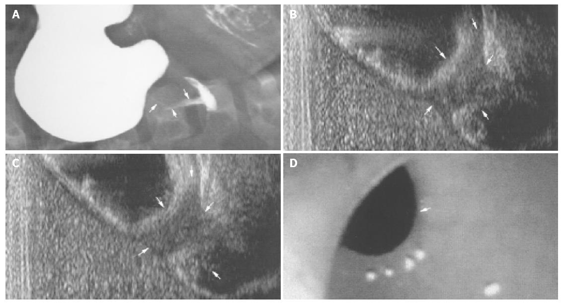
A 3-year-old boy was admitted with postprandial non-bilious vomiting and dull abdominal pain for 3 wk, and body weight loss of 2 kg. Blood-tinged vomitus was also occasionally noted. Physical examination revealed a slim boy with a body weight of 11.2 kg (<5 th percentile) and body height of 95.3 cm (60 th percentile). The otherwise physical examination was unremarkable. Laboratory studies including complete blood count, serum electrolytes, and liver function test were normal. Initial abdominal ultrasound (Acuson 128 XP/10 scanner, 7-MHz linear transducer, Mountain View, CA) revealed no lesions. Barium meal study showed a partial gastric outlet obstruction finding mimicking hypertrophic pyloric stenosis (Figure 1A). However, such an impression was not compatible with the clinical settings because a lack of the typical palpable “olive” in the right upper abdomen and the unusual late onset of presentation. Thus, abdominal ultrasound was repeated after water loading of the stomach by asking the patient to drink 500 mL of distilled water. An echogenic flap with an eccentric aperture (0.8 cm) at the gastric antral region and thickening of the distal antral wall beyond the flap were clearly demonstrated (Figure 1B). Turbulent flow around the aperture was noted during external compression of the upper abdomen reflecting the hindrance of water inflow through the hypertrophic distal antrum (Figure 1C). A thin jet-like flow through the pyloric canal to the duodenal bulb could also be identified. Subsequent gastric endoscopy (GIF N30, Olympus, Roswell, GA, USA) confirmed the presence of an antral web with an eccentric aperture (Figure 1D) and narrowed lumen beyond the web due to antral wall hypertrophy. Surgery confirmed the presence of an antral web with antral hypertrophy and prepyloric stenosis. Web excision and antropyloroplasty were performed. The patient recovered well and at the 24-mo follow-up, good body weight gain with a final weight of 20 kg (90 th percentile) and body height of 110 cm (75 th percentile) were noted.
The antral web or diaphragm is a thin septum, usually 2- to 4-mm thick located 1- to 7-cm from the pylorus. It typically projects into the gastric lumen perpendicular to the long axis of the antrum[1,2]. The etiology of the antral web is still controversial. It may originate from incomplete canalization of the foregut anlage during the 5-6th wk of the embryonic age, as an incomplete form of membranous atresia[1-3,8,9]. However, an acquired antral web in adults due to peptic diseases has also been documented[10]. The age of onset and clinical presentations of an antral web vary depending on the degree of obstruction and the size of its aperture, which may range from 2- to 30-mm [1-5]. Delays in diagnosis and treatment are not uncommon[2-5], and an aperture of less than 1 cm usually causes significant symptoms, as seen in our case.
Barium meal study allows accurate diagnosis of an antral web in 90% of cases[1-5]. The presence of a persistent, sharp band-like filling defect in the antral region as well as spraying of barium through a central or an eccentric aperture with a “jet effect” is the characteristic features. Distension of the antrum beyond the aperture may be seen, resembling the duodenal bulb and leading to the typical “double-bulb” appearance[1-5]. In this 3-year-old boy, the barium meal study demonstrated the imaging feature that was suggestive of hypertrophic pyloric stenosis which usually affects infants at the 2-4 wk of life. In retrospect, we realized that this misleading barium meal finding might be related to the eccentric location of the aperture of the antral web with distal antral muscular hypertrophy leading to luminal narrowing mimicking pyloric stenosis.
Diagnosis of an antral web can also be established with ultrasound[3,4,9]. Chew et al[9] proposed four ultrasound diagnostic criteria of an antral web, including demonstration of an echogenic diaphragm-like structure in the antral region, gastric dilatation, delay in gastric emptying, and a normal pylorus. In this particular case, the echogenic flap and eccentric aperture were clearly demonstrated on ultrasound after water-loading, but the antral chamber distal to the web remained poorly distended. Furthermore, turbulence was found at the aperture when intragastric fluid was forced to empty into the duodenal bulb during external compression of the distended stomach. This might have been due to the resistance to gastric outflow, plausibly ascribed to distal antral hypertrophy. In addition, instead of expanding the distal antral lumen, jet-like echogenic spraying distended the duodenal bulb, which indicated that the gastric fluid was being propelled through the narrowed antral lumen. To our knowledge, this is the first report of ultrasound diagnosis of an antral web associated with distal antral hypertrophy and prepyloric stenosis.
Endoscopy is helpful in confirming the presence of an antral web and in exploring other gastric pathologies such as peptic diseases, adhesion, and a heterotopic pancreas[1,2,8,11-13]. Endoscopic diagnostic criteria include a diaphragm with smooth mucosa and an opening of constant size, and normal peristalsis distal to the web[11]. In this small child, no evidence of peptic disease could be found on endoscopy. However, distal antral hypertrophy with poor peristalsis was noted confirming the ultrasound findings. We postulate that thickening of the distal antral wall could possibly be ascribed to reactive changes from the high-pressure inflow generated by the stomach through the aperture of the antral web. Nevertheless, the endoscope would be navigated through the distal antral lumen to the duodenal loop without difficulty once the antral web aperture was bypassed.
For a symptomatic antral web with gastric outlet obstruction, surgery remains the primary treatment method[1-5]. Most antral webs can be managed with a simple incision to excise the web[1,2]. Endoscopic transection or laser lysis of the web has also been described with satisfactory results[12,13]. However, accurate preoperative differentiation of an antral web with prepyloric stenosis from hypertrophic pylorus stenosis is important for surgical planning[5]. Hypertrophic pyloric stenosis can be managed by pyloromyotomy[5,6]. However, as in our patient, in addition to resection of the web, antropyloroplasty was also necessitated. With adequate distal antral lumen re-expansion, this small child enjoyed good recovery with satisfactory body weight and body height gain.
In summary, this report illustrates that an antral web with distal antral hypertrophy and prepyloric stenosis may mimic hypertrophic pylorus stenosis on a barium meal study.
Ultrasound study of the stomach after water loading with appropriate compression can offer a good anatomic delineation and an accurate preoperative diagnosis of this uncommon cause of gastric obstruction in a small child.
Assistant Editor Guo SY Edited by Wang XL
| 1. | Bell MJ, Ternberg JL, McAlister W, Keating JP, Tedesco FJ. Antral diaphragm--a cause of gastric outlet obstruction in infants and children. J Pediatr. 1977;90:196-202. [RCA] [PubMed] [DOI] [Full Text] [Cited by in Crossref: 38] [Cited by in RCA: 40] [Article Influence: 0.8] [Reference Citation Analysis (0)] |
| 2. | Bjorgvinsson E, Rudzki C, Lewicki AM. Antral web. Am J Gastroenterol. 1984;79:663-665. [PubMed] |
| 3. | Lui KW, Wong HF, Wan YL, Hung CF, Ng KK, Tseng JH. Antral web--a rare cause of vomiting in children. Pediatr Surg Int. 2000;16:424-425. [RCA] [PubMed] [DOI] [Full Text] [Cited by in Crossref: 20] [Cited by in RCA: 21] [Article Influence: 0.8] [Reference Citation Analysis (0)] |
| 4. | Van Winckel MA, Afschrift MB, Vande Walle JG. Ultrasound diagnosis of a prepyloric diaphragm. J Clin Ultrasound. 1994;22:141-143. [RCA] [PubMed] [DOI] [Full Text] [Cited by in Crossref: 5] [Cited by in RCA: 4] [Article Influence: 0.1] [Reference Citation Analysis (0)] |
| 5. | Mandell GA. Association of antral diaphragms and hypertrophic pyloric stenosis. AJR Am J Roentgenol. 1978;131:203-206. [RCA] [PubMed] [DOI] [Full Text] [Cited by in Crossref: 13] [Cited by in RCA: 13] [Article Influence: 0.3] [Reference Citation Analysis (0)] |
| 6. | Ferguson C, Morabito A, Bianchi A. Duodenal atresia and gastric antral web. A significant lesson to learn. Eur J Pediatr Surg. 2004;14:120-122. [RCA] [PubMed] [DOI] [Full Text] [Cited by in Crossref: 16] [Cited by in RCA: 16] [Article Influence: 0.8] [Reference Citation Analysis (0)] |
| 7. | Willcoxon RL, Farha GJ. Prepyloric antral stenosis. Report of a case with muscular hypertrophy and mucosal web. J Kans Med Soc. 1970;71:353-354. [PubMed] |
| 8. | Bell MJ, Ternberg JL, Keating JP, Moedjona S, McAlister W, Shackelford GD. Prepyloric gastric antral web: a puzzling epidemic. J Pediatr Surg. 1978;13:307-313. [RCA] [PubMed] [DOI] [Full Text] [Cited by in Crossref: 37] [Cited by in RCA: 37] [Article Influence: 0.8] [Reference Citation Analysis (0)] |
| 9. | Chew AL, Friedwald JP, Donovan C. Diagnosis of congenital antral web by ultrasound. Pediatr Radiol. 1992;22:342-343. [RCA] [PubMed] [DOI] [Full Text] [Cited by in Crossref: 13] [Cited by in RCA: 15] [Article Influence: 0.5] [Reference Citation Analysis (0)] |
| 10. | Huggins MJ, Friedman AC, Lichtenstein JE, Bova JG. Adult acquired antral web. Dig Dis Sci. 1982;27:80-83. [RCA] [PubMed] [DOI] [Full Text] [Cited by in Crossref: 10] [Cited by in RCA: 10] [Article Influence: 0.2] [Reference Citation Analysis (0)] |
| 11. | Banks PA, Waye JD. The gastroscopic appearance of antral web. Gastrointest Endosc. 1969;15:228-229. [PubMed] |
| 12. | Berr F, Rienmueller R, Sauerbruch T. Successful endoscopic transection of a partially obstructing antral diaphragm. Gastroenterology. 1985;89:1147-1151. [PubMed] |
| 13. | Al-Kawas FH. Endoscopic laser treatment of an obstructing antral web. Gastrointest Endosc. 1988;34:349-351. [RCA] [PubMed] [DOI] [Full Text] [Cited by in Crossref: 12] [Cited by in RCA: 14] [Article Influence: 0.4] [Reference Citation Analysis (0)] |


