INTRODUCTION
Multiseptate gallbladder is a very extremely rare congenital anomaly of the gallbladder. In the published cases, concomitant biliary diseases of the multiseptate gallbladder were reported as cholecystolithiasis, choledochal cyst, and hypoplasia. On the other hand, anomalous pancreaticobiliary ductal union is also one of the congenital anomalous biliary diseases and thought to be related with choledochal cyst or biliary tract malignancies. Along with the review of literature, reported here is a first patient of multiseptate gallbladder with anomalous pancreaticobiliary ductal union.
CASE REPORT
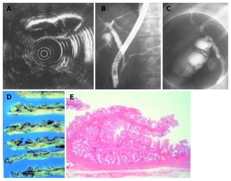
A 46-year-old Japanese female with gastric carcinoma underwent preoperative examinations at our institution. She had no complaints and her past history and family history were noncontributory. She reported no history of smoking and alcohol. Neither anemia nor jaundice was present. The abdomen was flat and soft. Liver, spleen, and mass could not be palpable. No lymph node swelling was palpated. Plain abdominal X-ray was normal. Laboratory data including tumor makers showed within normal range. Abdominal US and CT suspected septa in the gallbladder and showed neither cholecystolithiasis nor tumor lesion in the gallbladder. EUS demonstrated clearly multiple thin septa of the gallbladder (Figure 1A). ERCP revealed multiple faint septation in the neck portion of the gallbladder and combined with anomalous pancreaticobiliary ductal union. The common bile duct was not significantly dilated (Figures 1B and C).
Figure 1 A: Endoscopic ultrasonography of the gallbladder showing multiple thin septa in the neck portion.
B: Endoscopic retrograde cholangiopancreatography showing anomalous pancreaticobiliary ductal union. The common bile duct was not dilated. C: Endoscopic retrograde cholangiopancreatography showing multiple faint septation in the neck of the gallbladder. D:Longitudinal resected gallbladder shows multiple thin septa appearance at the neck portion. E: Microscopic findings of the resected gallbladder demonstrate the septa lined by normal typical columnar epithelium containing normal muscular layer (H&E stain, original magnification ×40).
Total gastrectomy and cholecystectomy were performed. In the macroscopic findings, the resected gallbladder was of normal size with smooth surface. Multiple thin septa were present only at the neck of the gallbladder. Neither tumor nor cholelithiasis was observed (Figure 1D). Microscopically, the septa were lined by typical columnar epithelium containing normal muscular layer of the gallbladder. There were mild chronic inflammation changes with the gallbladder. Pathological findings were consistent with multiseptate gallbladder (Figure 1E).
DISCUSSION
In recent years, congenital malformations of the gallbladder are increasingly diagnosed according to the rapid improvement of the imaging. The congenital malformations of the gallbladder are classified as anomalous forms, abnormal position, and absence. Multiseptate gallbladder is a very rare congenital anomaly form, which was first described by Simon and Tandon[1]. To our knowledge, only 30 cases including our case have been reported in the literature. A review of the cases described a female predominance; 21 of the patients were females. A mean age showed 29.4 years (range 3-70 years), and pediatric patients in four cases[2-5].
Most patients usually present with right upper abdominal pain, epigastralgia, and back pain, only six cases including our case were asymptomatic. Though the symptoms of multiseptate gallbladder patients were relieved after cholecystectomy, histological findings of the resected gallbladder in patients were usually of normal size with smooth surface and not always recognized as acute cholecystitis or cholecystolithiasis, and the exact mechanism of the biliary symptoms of multiseptate gallbladder is unknown. Saimura and co-workers investigated the mechanism of the symptoms of the patients with multiseptate gallbladder using biliary manometry and scintigraphy[6]. According to their study, both biliary manometry and scintigraphy indicated the impairment of the bile flow between the common bile duct and the gallbladder. One of the mechanism of the symptoms of multiseptate gallbladder is considered to be the impairment of the bile flow.
In nearly all literatures, pathological findings of multiseptate gallbladder reveal numerous septa, which consisted of a normal muscularis layer of the gallbladder wall, like a honeycomb appearance. Though the septa usually existed in the whole of the gallbladder, in some cases, the septations were confined to the neck, the body and the fundus alone[2,7,8]. This case presented the multiple septa in the neck portion of the gallbladder.
To make a diagnosis of multiseptate gallbladder exactly, it is important to detect clearly the septa in the gallbladder. Diagnostic imaging modalities for this disease were reported as abdominal US, abdominal CT[9] ERCP[7,10] oral cholecys-tography, and 99mTc hepatobiliary imaging[11]. In some asymptomatic cases, the diagnosis were made incidentally by abdominal US[3,4,8,12]. This case presented more clearly multiple thin septa in the neck portion of the gallbladder on EUS rather than other imagings. EUS is considered to be a very useful modality for the diagnosis of the cases with partial multiseptation.
Concomitant biliary disease of the patients with multiseptate gallbladder besides our case was reported in six patients; cholelithiasis in two patients[3,13] choledochal cyst in two[5,14] and hypoplasia of the gallbladder in two[15,16]. No cases that coexisted with malignancies were reported in the literature. On the other hand, anomalous pancreaticobiliary ductal union is also known as the congenital malformation of the pancreaticobiliary duct system and is thought to be related to choledochal cyst and biliary tract carcinoma. Though anomalous pancreaticobiliary ductal union is frequently observed in patients with congenital choledochal cyst, the cases without choledochal cyst are recognized to have great malignant potential of the gallbladder[17,18]. In this case, ERCP demonstrated anomalous pancreaticobiliary ductal union without choledochal cyst.
Our case is asymptomatic and has the septations at the only neck of the gallbladder. Besides these unique findings, this present case is the first report of multiseptate gallbladder associated with anomalous pancreaticobiliary ductal union in the world literature. To clarify more characters of the multiseptate gallbladder, examination of a larger patient population will be needed and further studies will be required.


