Published online Sep 21, 2005. doi: 10.3748/wjg.v11.i35.5517
Revised: August 27, 2004
Accepted: August 30, 2004
Published online: September 21, 2005
AIM: To determine the role of acupuncture therapy in treating experimental gastric ulcer in rats.
METHODS: Twenty-eight adult male Sprague-Dawley rats were randomly divided into four groups (pre-acupuncture group; acupuncture group; paradistance-acupuncture group; and control group), and pre-acupuncture, paradistance-acupuncture, and control groups received 5 μL acetic acid (200 mL/L HAc) injection after a same course of electroacupuncture (EA) treatment (4 Hz, 0.6 mA, 0.45 ms, 45 min for 4 d). The rats in these three groups recovered within 4 d. The acupuncture group received EA therapy for 4 d, after HAc injection. The stomach was dissected to compare the pathological structures of ulcer. Also c-Fos activation in the nuclei of solitary tract (NTS) was observed under microscope after regular immunohistochemistry staining of brain stem sections.
RESULTS: The number of ulcers was different among the four groups, especially between control group and paradistance-acupuncture group or pre-acupuncture group. In the latter group, the number of ulcers was much less. The gastric ulcer area was consistent with the histopathological results, indicating that pre-acupuncture had an obvious therapeutic effect on gastric ulcers. Acupuncture had a very modest effect and paradistance-acupuncture had no effect on gastric ulcers. No therapeutic effect was found in the control group. Fos-Li neurons in NTS induced by noxious gastric ulcer showed a significant difference between pre-acupuncture and control groups.
CONCLUSION: Acupuncture before ulceration can obviously alleviate ulcer. The production of c-Fos proves that the vagus nerve mediates the induction of c-Fos in nuclei of solitary tract following experimental ulceration, suggesting that parasympathetic afferents promote the process of noxious visceral stimulation.
- Citation: Wang H, Wang CY, Zhang JS, Sun L, Sun JP, Tian QH, Jin XL, Yin L. Acupuncture therapy for experimental stomach ulcer and c-Fos expression in rats. World J Gastroenterol 2005; 11(35): 5517-5520
- URL: https://www.wjgnet.com/1007-9327/full/v11/i35/5517.htm
- DOI: https://dx.doi.org/10.3748/wjg.v11.i35.5517
Acupuncture has been used for the treatment of a wide variety of conditions[1] including fever, inflammation and infection, especially stomach diseases. But the efficacy and mechanism of acupuncture in prevention of acid-induced ulcers are unknown. Here the previous animal ulceration model[2] by injection of acetic acid (HAc) was used to study the effectiveness of acupuncture. The zusanli (St36) acupuncture was chosen, because it may have therapeutic functions for stomach diseases and it may be related with the anti-inflammation effect of vagus nerve[3]. C-Fos has been used as a marker for activity in the central nervous system following noxious somatic and visceral stimulation. Experimental gastric ulcer could induce significant c-Fos activity in the nuclei of the solitary tract (NTS) and lumbosacral spinal cord, which receives more parasympathetic afferent inputs than sympathetic afferent inputs.
The study was carried out to examine the effect of electroacupuncture (EA) on attenuating the inflammation of animals subjected to acid-induced gastric ulcer. The acupuncture effect on gastric ulcer was compared among different groups and their relations with c-Fos-activated neurons in vagus-mediated nuclear NTS were analyzed.
Twenty-eight adult male Sprague-Dawley rats weighing 150-250 g were included in this study. The rats were divided into four groups randomly and housed in a 12-h light and dark cycle with free access to food and water. They were deprived of food but not water 12 h prior to experiment. The experiment was approved by Chinese Animal Committee.
Among several models[4,5], a previously established model to induce gastric ulcer in rats was used. The rats were anesthetized with 45-50 mg/kg pentobarbital intraperitoneally. A midline incision was made to expose the stomach, and 5 μL of 200 mL/L HAc in sterile saline was injected into the subserosal layer of the dorsal and ventral stomach wall and then the epigastric incision was closed. After a recovery period of 4 d, just before the repair time for ulcer[6,7], EA was performed for the acupuncture group. Then the animals were anesthetized, the stomach was opened along the greater curvature, and then pinned to a corkboard. Macroscopic mucosal damage and gastric ulcer were immediately evaluated and gastric ulcer tissues were stained with hematoxylin and eosin (HE). The rat body was perfused immediately after the stomach was removed.
EA treatment (4 Hz, 0.6 mA, 0.45 ms) was carried out for 45 min/day for 4 consecutive d prior to acid injection. The rats were anesthetized, EA was applied with stainless steel needles inserted bilaterally into points corresponding to zusanli (St 36) in human beings. Since EA stimulation produced slight muscle twitch, and did not raise the blood pressure of the anaesthetized animals, it was mostly an innocuous stimulation.
Gastric mucosal damage was quantitated by measuring the area and the number of mucosal lesions. The length and width of each gastric lesion were measured to assess the area of mucosal damage and then the total area of superficial ulceration was calculated. The area of aggregate lesions was expressed as square millimeters.
Under deep sodium pentobarbital anesthesia, the animals were perfused transcardially with 100 mL saline, followed by 500 mL fixative containing 40 g/L paraformaldehyde in 0.1 mol/L phosphate buffer (PB, pH 7.4). The brain tissue containing the medulla oblongata was removed and stored in 300 g/L sucrose (4 °C) until sedimentation of the blocks, and cut into serial 30 μm coronal sections on a cryostat. For anti-Fos immunostaining, the sections were treated with methanol, H2O2 and 3 g/L Triton X-100 for 30 min, then successively incubated with polyclonal primary rabbit anti-Fos antibody (1:10 000, Sigma) for 16 h, donkey anti-rabbit antibody (1:200, Vector) for 3 h, mixture complex liquid of avidin-biotin(1:100, Vector) for 1 h at room temperature. Then the sections were visualized by 0.5 g/L diaminobenzidine(DAB) and 0.3 g/L H2O2 (DAB kit, Dako) in distilled water, and rinsed in phosphate buffered saline (PBS) for 10 min thrice after each step. One set of sections were stained with Nissle to get the comparative nuclei in brain stem sections.
Results were expressed as mean±SD. The analysis of variance (ANOVA) was employed for statistical evaluations and comparisons of the data. The differences in mean values between two experimental g roups were evaluated by t-test. P<0.05 was considered statistically significant.
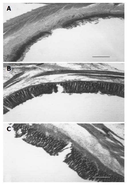
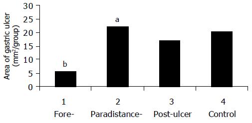
Significantly histological changes were observed after submucosal injection of HAc into the 4 groups. Microscopic examination showed that gastric ulcers induced by HAc-injection in acupuncture group had much more repairing area of submucosa than the untreated rats (Figure 1), and the whole area of gastric ulceration was found in the former group (P<0.01, Table 1). The ulceration area of paradistance acupuncture group increased obviously at the same time. The measured results were 5.6±0.5 mm2 in early acupuncture prior to ulcer, 22.1±5.9 mm2 in paradistance acupuncture group, and 16.90.8 mm2 in acupuncture after ulceration. The submucosa of stomach in rats subjected to early stage acupuncture was well protected, but acupuncture therapy after HAc-injection failed to prevent mucosal ulcer injury (P>0.1, Figure 2). In comparison to the area of gastric ulceration, the acupuncture-dependent inhibition of ulceration could be observed (Figure 2).
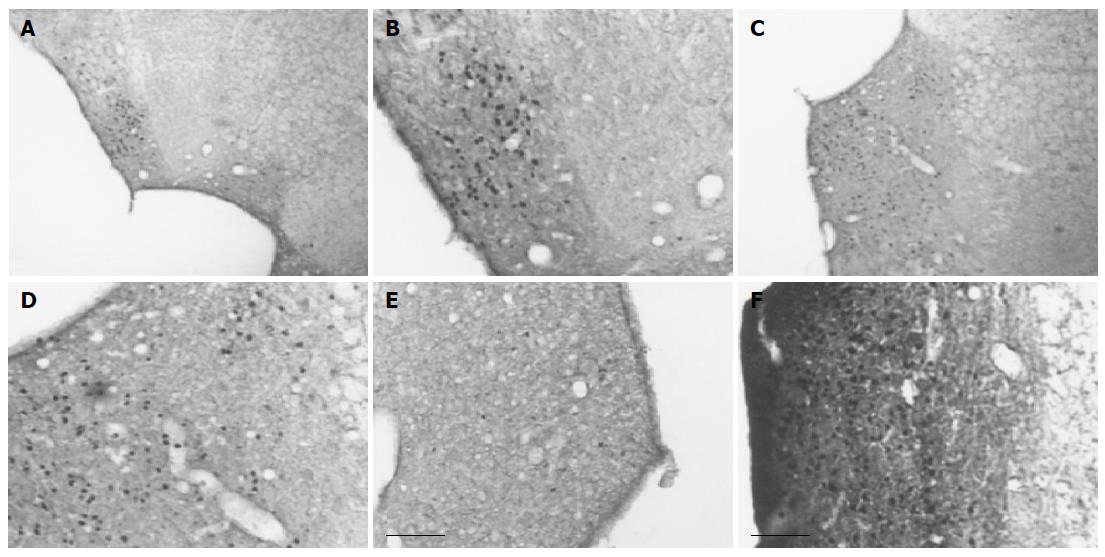
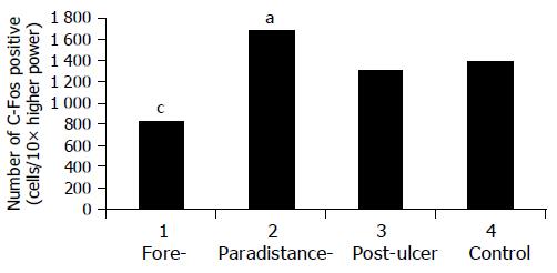
Most Fos-Li neurons were distributed mainly around NTS (Figures 3: B, D, F). There were also some sparsely distributed Fos-Li neurons in surrounding area. At the lower (Figures 3: A, C) or higher magnification (Figures 3: B, D, E, F), the high density of Fos-Li neurons was obviously shown in non-acupuncture group (Figure 3D) and paradistance group (Figure 3F). The acupuncture therapy in early stage could prevent the production of c-Fos (Figure 3E). The number of c-Fos-stained nuclei was counted from NTS at three levels of brain stem in each group (seven rats) animals. For each section three fields were chosen to count the Fos-Li expressing neurons. In pre-acupuncture group the induced Fos-Li neurons were fewer (Figure 4, P<0.05). In both paradistance acupuncture group (Figure 4, P<0.05) and acupuncture group, the number of Fos-Li neurons significantly increased (Figure 4). In the control group the number of Fos-Li neurons was also higher (Figure 4).
In our study, the acupuncture point was accurate and reasonable. When the rats were anesthetized, EA was applied with stainless steel needles inserted bilaterally into points corresponding to zusanli (St36) in human beings. EA stimulation produced slight muscle twitch, but did not raise the blood pressure in anaesthetized animals, indicating that it is an innocuous stimulation. Fos-Li cells induced by these factors should die after so long a repair time and also some of them might spread to other brain areas especially in the latter situation. It means that even if acupuncture and surgery could induce c-Fos protein expression, but it could be neglected over the long enough recovery time. The results are consistent with the hypotheses, otherwise sham group should have had fewer c-Fos-positive neurons than acupuncture group. But the opposite results we obtained indicate that the ulceration in control group can induce more noxious signals from ulcer inflammation than the acupuncture-treated animals.
HAc injected into the gastric wall induced significant inflammation and ulceration in rats. Four days were chosen for the ulceration time, because this is the most possible period for inducing ulceration[4,5]. Pathological examination showed that the acupuncture (St36) attenuated gastric ulcer in experimental rats. The repair in submucosa was more complete than that in non acupuncture group (Figures 1 and 3). The number and area of ulceration were different (Table 1).
Acupuncture has been used to treat gastrointestinal symptoms in China for more than 3 000 years. However, the mechanism of its beneficial effects remains unknown. It was reported that acupuncture at the right lower abdomen causes a transient relaxation of the stomach, and acupuncture-induced gastric relaxations can be abolished by guanethidine, propranolol, splanchnic ganglionectomy, spinal cord transection, and spinomedullary transection[8]. In contrast, N (G)-nitro-L-arginine, phentolamine, truncal vagotomy, and pontomedullary transection have no effect. Popova et al[9], have proved the effect of acupuncture on activity of antioxidant system in rats with gastroduodenal ulcer. More recently the effect of acupuncture on young pigs with enteropathogenic Escherichia coli diarrhea has been evaluated by staining with hematoxylin and eosin[10]. All these results indicate that acupuncture treatment is effective in controlling some stomach diseases at their early stage.
Concerning both of these studies and our results, the number of c-Fos immunopositive cells at the NTS in medulla was changed because of prevailing acupuncture. NTS receives gastrointestinal mechno- and chemosensory information via the vagus nerve and vagal afferents may be involved in chemonoreception, which means that pain signals induced by gastric ulcer can be traced by the most related nucleus-NTS in medulla and this effect can be deduced by accurate acupuncture via vagus nerve, suggesting that acupuncture-induced gastric ulcer reduction is mediated via the somato-parasympathetic reflex system. But acupuncture afferent limb is composed of abdominal cutaneous and muscle afferent nerves. Though the direct or indirect efferent limb is questionable, our hypothesis is via the gastric parasympathetic nerve and the reflex center is within the medulla. NTS neurons may play an important role in mediating this reflex and are the 慴rain of the gut for autonomic control of digestion. Because 88-90% of axons in the vagus nerves are afferent nerve fibers that project to the nucleus tractus solitaries and area postrema[11,12], acupuncture prevents mainly brain structures from formation and increase of the products of Fos-Li neurons, suggesting that vagus nerve mediates induction of c-Fos in NTS following experimental ulceration, and that parasympathetic afferents promote noxious visceral stimulation. Results from this study are in agreement with some reports[13].
Although there is evidence that induction of Fos-Li neurons does not depend on the integrity of vagus nerves[8,14]. Tada et al[8], suggested that acupuncture treatment of gastric ulcer can be mediated via the somatosympathetic reflex system. Its afferent limb is composed of abdominal cutaneous and muscle afferent nerves. Its efferent limb is the gastric sympathetic nerve and the reflex center is within the ventral lateral medulla. Our present study strongly supports that vagus nerve itself plays a very important role in peripheral gastric organ and central brain communication and cytokine-brain communication[15], because ulcer itself is a kind of cytokine-induced inflammation. The hypothesis that inflammation of gastric ulcer activates afferent vagal fibers that terminate in the nucleus tractus solitarius and may be adjusted by early stage acupuncture has been proved. Previous studies showed that vagal nerve may be a neural route and vagotomy blocks the afferent signaling to the central nerve system and the effects of peripheral cytokines. Peripheral cytokines induced by ulcer inflammation may influence the central nerve system by blood-borne routes of communication.
In conclusion, our study provides the possible neural mechanism of acupuncture underlying gastric ulcer formation. NTS neurons may play an important role in mediating gastric ulcer.
Science Editor Wang XL, Zhu LH and Guo SY Language Editor Elsevier HK
| 1. | Pei WF, Xu GS, Sun Y, Zhu SL, Zhang DQ. Protective effect of electroacupuncture and moxibustion on gastric mucosal damage and its relation with nitric oxide in rats. World J Gastroenterol. 2000;6:424-427. [PubMed] |
| 2. | Takahashi S, Shigeta J, Inoue H, Tanabe T, Okabe S. Localization of cyclooxygenase-2 and regulation of its mRNA expression in gastric ulcers in rats. Am J Physiol. 1998;275:G1137-G1145. [PubMed] |
| 3. | Borovikova LV, Ivanova S, Zhang M, Yang H, Botchkina GI, Watkins LR, Wang H, Abumrad N, Eaton JW, Tracey KJ. Vagus nerve stimulation attenuates the systemic inflammatory response to endotoxin. Nature. 2000;405:458-462. [RCA] [PubMed] [DOI] [Full Text] [Cited by in Crossref: 2722] [Cited by in RCA: 2969] [Article Influence: 118.8] [Reference Citation Analysis (0)] |
| 4. | Sisco M, Mustoe TA. Animal models of ischemic wound healing. Toward an approximation of human chronic cutaneous ulcers in rabbit and rat. Methods Mol Med. 2003;78:55-65. [PubMed] |
| 5. | Li Z, Shi A, Karlsson FA. Transient increase of rat gastric amylin in the neonatal period and in experimental ulcers. J Gastroenterol. 2002;37:172-176. [RCA] [PubMed] [DOI] [Full Text] [Cited by in Crossref: 3] [Cited by in RCA: 3] [Article Influence: 0.1] [Reference Citation Analysis (0)] |
| 6. | Bielefeldt K, Ozaki N, Gebhart GF. Experimental ulcers alter voltage-sensitive sodium currents in rat gastric sensory neurons. Gastroenterology. 2002;122:394-405. [RCA] [PubMed] [DOI] [Full Text] [Cited by in Crossref: 84] [Cited by in RCA: 77] [Article Influence: 3.3] [Reference Citation Analysis (0)] |
| 7. | Pawlik M, Ptak A, Pajdo R, Konturek PC, Brzozowski T, Konturek SJ. Sensory nerves and calcitonin gene related peptide in the effect of ischemic preconditioning on acute and chronic gastric lesions induced by ischemia-reperfusion. J Physiol Pharmacol. 2001;52:569-581. [PubMed] |
| 8. | Tada H, Fujita M, Harris M, Tatewaki M, Nakagawa K, Yamamura T, Pappas TN, Takahashi T. Neural mechanism of acupuncture-induced gastric relaxations in rats. Dig Dis Sci. 2003;48:59-68. [RCA] [PubMed] [DOI] [Full Text] [Cited by in Crossref: 62] [Cited by in RCA: 63] [Article Influence: 2.9] [Reference Citation Analysis (0)] |
| 9. | Popova SP. Effect of acupuncture on dynamics of changes in lipid peroxidation and activity of antioxidant system in rats with gastroduodenal ulcer. Fiziol Zh. 2002;48:54-59. [PubMed] |
| 10. | Park ES, Jo S, Seong JK, Nam TC, Yang IS, Choi MC, Yoon YS. Effect of acupuncture in the treatment of young pigs with induced Escherichia coli diarrhea. J Vet Sci. 2003;4:125-128. [PubMed] |
| 11. | Holzer P, Michl T, Danzer M, Jocic M, Schicho R, Lippe IT. Surveillance of the gastrointestinal mucosa by sensory neurons. J Physiol Pharmacol. 2001;52:505-521. [PubMed] |
| 12. | Berthoud HR, Neuhuber WL. Functional and chemical anatomy of the afferent vagal system. Auton Neurosci. 2000;85:1-17. [RCA] [PubMed] [DOI] [Full Text] [Cited by in Crossref: 768] [Cited by in RCA: 821] [Article Influence: 32.8] [Reference Citation Analysis (0)] |
| 13. | Furness JB, Clerc N, Kunze WA. Memory in the enteric nervous system. Gut. 2000;47 Suppl 4:iv60-iv2; discussion iv76. [PubMed] |
| 14. | Hermann GE, Emch GS, Tovar CA, Rogers RC. c-Fos generation in the dorsal vagal complex after systemic endotoxin is not dependent on the vagus nerve. Am J Physiol Regul Integr Comp Physiol. 2001;280:R289-R299. [PubMed] |
| 15. | Ge X, Yang Z, Duan L, Rao Z. Evidence for involvement of the neural pathway containing the peripheral vagus nerve, medullary visceral zone and central amygdaloid nucleus in neuroimmunomodulation. Brain Res. 2001;914:149-158. [RCA] [DOI] [Full Text] [Cited by in Crossref: 19] [Cited by in RCA: 17] [Article Influence: 0.7] [Reference Citation Analysis (0)] |


