Published online Sep 7, 2005. doi: 10.3748/wjg.v11.i33.5251
Revised: March 18, 2005
Accepted: March 21, 2005
Published online: September 7, 2005
A 60-year-old female patient suffered unhealed wounds over left flank for around 30 years after surgical removal of left renal stones. Fecal material spilled from the two small openings of the scar, bothered her all day long. During the course of the 30 years, she suffered from intermittent fever, diarrhea and wound pain and presented with malnourished condition. After serial examinations, tumor associated with iatrogenic colo-cutaneous fistula was impressed and she received en bloc resection. Pathology revealed squamous cell carcinoma arising from the fistula with colon and spleen invasion. To the best of our knowledge, no such case has been reported, as yet.
- Citation: Lee YT, Hsu SD, Kuo CL, Chou DA, Lin MS, Huang MH, Wu HS. Squamous cell carcinoma arising from longstanding colocutaneous fistula: A case report. World J Gastroenterol 2005; 11(33): 5251-5253
- URL: https://www.wjgnet.com/1007-9327/full/v11/i33/5251.htm
- DOI: https://dx.doi.org/10.3748/wjg.v11.i33.5251
Malignant tumors arising from previously existing fistulas are rare. Squamous cell carcinoma arising from colocutaneous fistula has never been reported. Herein, we report a case of squamous cell carcinoma arising from previously existing longstanding colocutaneous fistula. The diagnosis made has been based on high suspicion, history, imaging and pathology.
A 60-year-old female patient was admitted due to general weakness, anorexia accompanied with fever and chills for 2 wk. She denied any other systemic disease. The patient originally was diagnosed with left renal stones and had left nephrectomy, at some other hospital three decades ago. She got stool spillage from surgical wound on left flank after that surgery. Despite wound treatment, two fistulous openings were left with intermittent fecal discharge. During this period, she received supportive treatment while infective symptoms such as fever, chills, diarrhea and local cellulites were present.
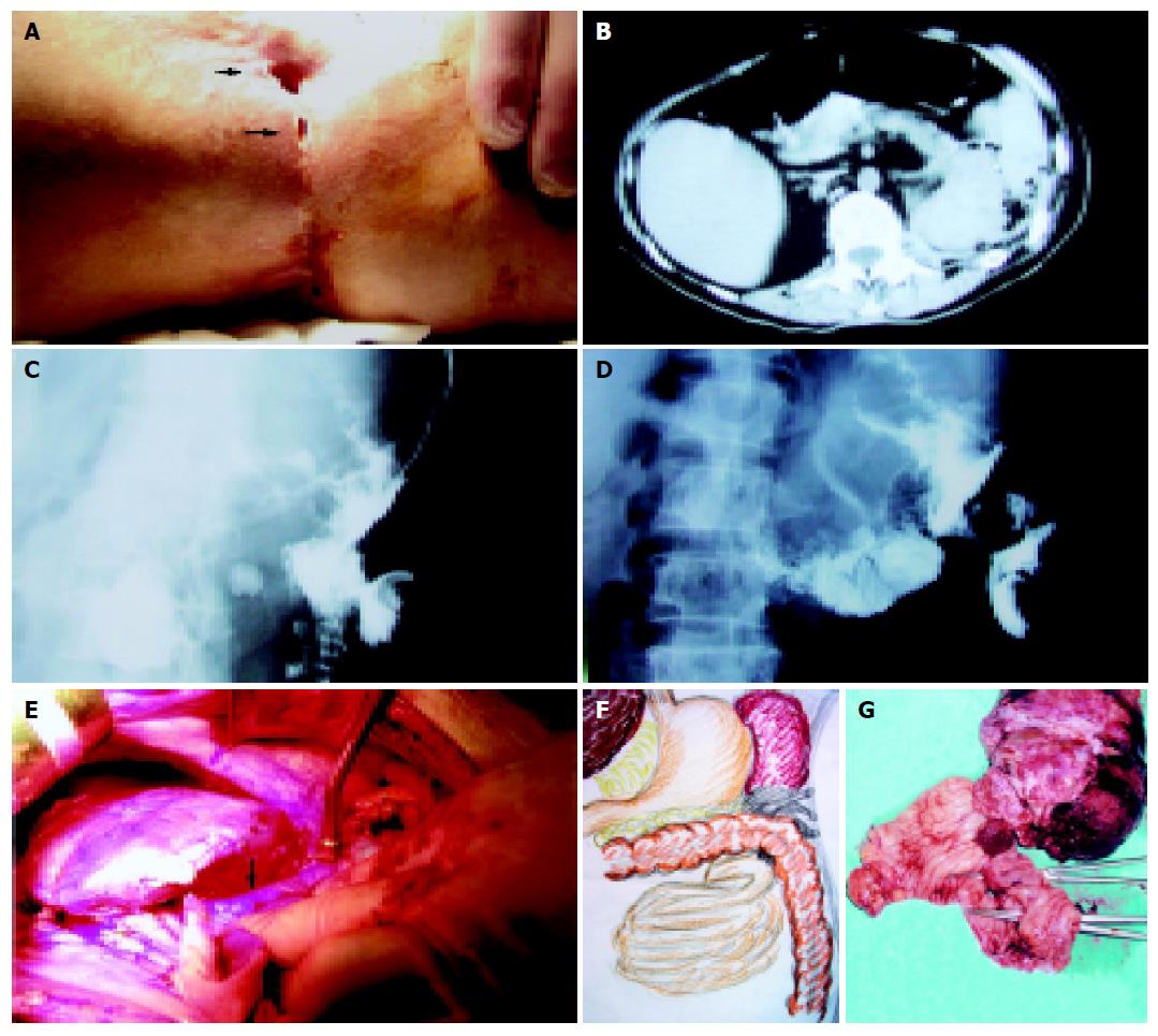
We noted that she was a cachectic, frail female with pale conjunctivae. The abdomen was palpated without tenderness and the bowels were normally peristaltic on auscultation. Two chronic unhealing wounds involving the left flank with fecal discharge were noted (Figure 1A). Local erythematous, swollen and tender to palpated were noted. No enlarged lymph nodes were palpated. Anemia (Hb 4.4 g/dL) and chronic renal insufficiency (Cr 2.3 mg/dL) were noted. Abdominal computed tomography discovered splenomegaly with soft tissue density adjacent to it and the splenic flexure of T-colon with air bubbles (Figure 1B). After infection had subsided, fistulograms revealed communication between skin and bowel tract (Figures 1C and D). Under a period of nutritional support, surgical intervention was performed. At laparotomy, the low pole of the spleen adhered to the splenic flexure of T-colon densely with irregular soft tissue beside the region (Figure 1E). Partial wall of the jejunum, 10 cm distal to Treitz’s ligament, adhered to the distal T-colon was noted (Figure 1F). En bloc resection with splenectomy, segmental resection of colon with primary anastomosis and wedge resection of jejunum were performed (Figure 1G). Postoperative course was grossly smooth. She is being followed up at the outpatient department.
The pathologist reported moderately differentiated squamous cell carcinoma in virtually all specimens (Figure 2A). However, the microscopic photographs revealed the tumor cells arising from the fistulas (Figure 2B) with spleen (Figure 2C) and colon invasion (Figure 2D).
It is well-known that surgery is still the most common cause of entero-cutaneous fistula. The causes of persistent entero-cutaneous fistula include foreign body, radiation, infection, inflammation, epithelization, neoplasm and distal obstruction[1]. Squamous cell carcinoma can develop from chronic ulcers, scars, wounds, sinuses, and fistulas[2]. The latent periods are long and take around 37 years for patients with burn scars except 1-7 years for immunocompromised patients[2,3]. The most significant factor reported in predicting the outcome for the squamous cell carcinoma from the pre-existing scar or sinus was the grade of the tumor[4]. Squamous cell carcinoma associated with prior renal stones have always been reported[5] and the median survival time was 3.6 mo[6]. It was dismal and was not compatible with the long-term history of the patients. We might consider the development of squamous cell carcinoma as the result of chronic irritation and infection due to unhealed wounds[7]. The strong evidence was that the microscopic photographs revealed the origination of tumor cells from the epithelium of the fistulous tract. However, there are no prior reported articles available as this case. Because of its insidious course, the long-standing colo-cutaneous fistula should be examined carefully for tumor development. The early nutritional intervention is important for patients with entero-cutaneous fistula[8] and surgery is inevitable for long-term unhealed fistula.
Science Editor Guo SY Language Editor Elsevier HK
| 1. | Courtney M, Townsend R Jr. Daniel Beauchamp B, Mark Evers, Kenneth L. Mattox. Sabinston Textbook of Surgery 16th ed. 2001;218-219. |
| 2. | Sarani B, Orkin BA. Squamous cell carcinoma arising in an unhealed wound in Crohn's disease. South Med J. 1997;90:940-942. [RCA] [PubMed] [DOI] [Full Text] [Cited by in Crossref: 8] [Cited by in RCA: 12] [Article Influence: 0.4] [Reference Citation Analysis (0)] |
| 3. | Edwards MJ, Hirsch RM, Broadwater JR, Netscher DT, Ames FC. Squamous cell carcinoma arising in previously burned or irradiated skin. Arch Surg. 1989;124:115-117. [RCA] [PubMed] [DOI] [Full Text] [Cited by in Crossref: 93] [Cited by in RCA: 83] [Article Influence: 2.3] [Reference Citation Analysis (0)] |
| 4. | Lifeso RM, Rooney RJ, el-Shaker M. Post-traumatic squamous-cell carcinoma. J Bone Joint Surg Am. 1990;72:12-18. [PubMed] |
| 5. | Ito H, Oishi M, Murase T. A case of squamous cell carcinoma of the renal pelvis originating from a functional solitary kidney associated with renal stones. Hinyokika Kiyo. 1988;34:2171-2174. [PubMed] |
| 6. | Raghavendran M, Rastogi A, Dubey D, Chaudhary H, Kumar A, Srivastava A, Mandhani A, Krishnani N, Kapoor R. Stones associated renal pelvic malignancies. Indian J Cancer. 2003;40:108-112. [PubMed] |
| 7. | Mosavy SH, Tayebi SA. Carcinoma arising in a perianal sinus tract: report of a case. Dis Colon Rectum. 1975;18:416-417. [RCA] [PubMed] [DOI] [Full Text] [Cited by in Crossref: 1] [Cited by in RCA: 2] [Article Influence: 0.0] [Reference Citation Analysis (0)] |
| 8. | Wang XB, Ren JA, Li JS. Sequential changes of body composition in patients with enterocutaneous fistula during the 10 days after admission. World J Gastroenterol. 2002;8:1149-1152. [PubMed] |


