INTRODUCTION
Gastric epithelial polyps are uncommon, estimated to be present in less than 2% of all endoscopic examinations[1]. Up to 90% of all gastric polyps are benign, can usually be classified as inflammatory, regenerative or hyperplastic, and are often less than 1 cm in size[2,3]. Only 5-10% of all gastric polyps are adenomas. Malignant change occurs in less than 1%, usually only in those that are over 1 cm[4].
Since the vast majority of gastric polyps are not neoplastic, and have a very low risk of containing a carcinoma, some authorities have argued that polyp biopsy alone may be sufficient; indeed, most adenomas are reported to display a maximal degree of dysplastic change in the most superficial portion of the polyp permitting biopsy detection[5]. Polyps may also cause symptoms related to bleeding or intermittent outlet obstruction, and may be removed endoscopically or at surgical resection. However, in some centers, even larger asymptomatic polyps may be biopsied, rather than excised, especially in elderly patients[5]. It has been argued that polypectomy is not without significant risk, particularly from hemorrhage after gastric polypectomy where risk has been historically estimated to be as high as 4%[5,6].
Gastric adenomas are usually solitary and asymptomatic. They occur primarily in the antrum and their frequency increases with aging[4]. Logically, gastric adenomas are best completely excised, so that the malignant potential, although very low, is removed. If endoscopic removal is not possible, then surgical resection might be considered, but then the individual risk and benefits must be weighed compared to long-term endoscopic monitoring, since it has been estimated that about 11% of gastric adenomas develop carcinoma within 4 years[4]. Unless the polyps cause symptoms, however, some have advocated that it may be reasonable to do nothing[5].
The present case was unusual in that a prolapsing adeno-matous gastric polyp, complicated by an adenocarcinoma, was responsible for gastric outlet obstructive symptoms and these were completely resolved with endoscopic excision of the gastric polyp.
CASE REPORT
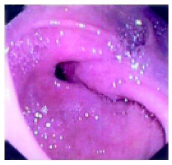
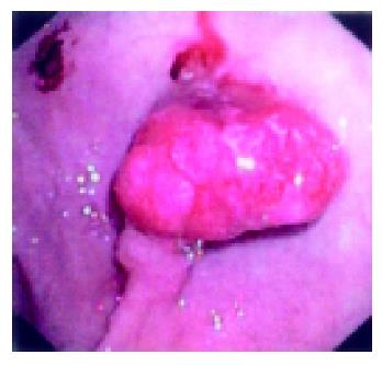
A 69-years old Indo-Canadian male with chronic lymphocytic leukemia was referred by his hematologist for investigation of a progressively developing iron deficiency anemia over the prior year. He felt occasional fullness and nausea after meals. There were no other symptoms. He denied hematemesis, rectal bleeding, melena and occult blood testing was negative. Except for a single pea-sized cervical lymph node, there was no other lymphadenopathy. There was no hepatomegaly or splenomegaly. Laboratory studies revealed a hemoglobin of 109 g/L (normal, 135-175) and a white blood cell count of 13.4×A9/L (normal, 4.0-11.0), the majority being lymphocytes. Serum ferritin was 3.1 ug/L (normal, 20-300). INR and PTT were normal. Prior studies, including flow cytometry, had confirmed the typical immunophenotype of chronic lymphocytic leukemia. Upper endoscopic evaluation showed a large polyp with a stalk originating in the gastric antrum and prolapsing through the pylorus (Figure 1). After the polyp was maneuvered into the stomach, it was noted to be irregular with evidence of surface friability and ulceration (Figure 2). After the polyp was excised by snare polypectomy, pathological evaluation was reported to show an adenomatous polyp with high grade dysplasia along with foci of intramucosal carcinoma. Additional sections through the base of the polyp stalk, however, confirmed that polyp excision was complete without evidence of invasion into the submucosa. No malignant cells were detected in the polyp stalk or cut margin. Other gastric biopsies were normal and stains for Helicobacter pylori were negative.
Figure 1 Polypoid lesion with long stalk protruding through the pylorus into the duodenum.
Figure 2 Slightly irregular gastric antral polyp with eroded surface.
Polypectomy specimen showed adenoma with high-grade dysplasia and invasive intramucosal carcinoma. No invasion into stalk or submucosa.
DISCUSSION
This patient presented with a large gastric antral polyp that had prolapsed through the pylorus, causing symptoms due to a gastric outlet obstruction. The polyp was endoscopically excised and proved to be a large gastric adenoma com-plicated by adenocarcinoma. This is distinctly unusual. Endoscopic removal or excision of symptomatic and prolapsing gastric polyps have been occasionally reported in the literature, however, these have been, as might be anticipated, primarily large, sometimes pedunculated polyps with inflammatory, regenerative or hyperplastic histopa-thologic features, rather than adenomas or adenomas with superimposed carcinomas[7-12].
In the earlier endoscopic literature, endoscopic biopsy alone for most lesions was thought to be sufficient, and if the histopathologic changes in these biopsies were consistent with an adenoma, then endoscopic polypectomy, or surgical resection, could be considered[13-16]. Later, endoscopic excision, rather than biopsy alone, was recommended since the procedure could be done safely and since foci of in situ malignant change could be detected in some adenomas. Others have also demonstrated the detection of carcinoma or dysplastic change in hyperplastic gastric polyps[17-19]. Given the evolution of endoscopic accessories for control of post-polypectomy bleeding vessels (e.g., loops, clips), the excision of large, especially solitary, gastric polyps has clearly been facilitated.
An additional management issue for the future was also considered here. Gastric polyps that have been completely excised and contain invasive cancer may require additional evaluation. Data is currently very limited for gastric polyps on the role of a further local surgical resection to examine adjacent lymph nodes for metastases. It appears that the risk of lymph node metastases is exceedingly low, but it is not zero[20]. Previous studies of so-called early gastric cancer confined to the mucosa or submucosa may provide some guidance. For early gastric cancers of the polypoid subtype (type 1) treated with surgical resection, 1.8% of patients with intramucosal carcinoma had nodal metastases compared to 18.5% with submucosal involvement[5]. Thus, until more data is available, it would appear reasonable to treat all polyps with pathologically-defined intramucosal carcinoma alone with endoscopic resection alone, while an additional surgical resection should be considered, if submucosal infiltration is present.
Science Editor Guo SY Language Editor Elsevier HK


