Brief Reports Open Access
Copyright ©2005 Baishideng Publishing Group Inc. All rights reserved.
World J Gastroenterol. Jun 14, 2005; 11(22): 3485-3489
Published online Jun 14, 2005. doi: 10.3748/wjg.v11.i22.3485
Comparative proteome analysis of untreated and Helicobacter pylori-treated HepG2
Yan Zhang, Xue-Gong Fan, Ren Chen, Xue-Fei Tian, Department of Infectious Diseases, Xiangya Hospital, Central South University, Changsha 410008, Hunan Province, China
Zhi-Qiang Xiao, Xue-Ping Feng, Zhao-Hui Chen, Medical Research Center, Xiangya Hospital, Central South University, Changsha 410008, Hunan Province, China
Author contributions: All authors contributed equally to the work.
Correspondence to: Xue-Gong Fan, Department of Infectious Diseases, Xiangya Hospital, Central South University, Changsha 410008, Hunan Province, China. xgfan@hotmail.com
Telephone: +86-731-4327392 Fax: +86-731-4327332
Received: July 2, 2004
Revised: July 3, 2004
Accepted: September 30, 2004
Published online: June 14, 2005

Abstract

AIM: To investigate the pathological effect of Helicobacter pylori (H pylori) on human hepatic cells, proteomic methods were used to find and to identify proteins that were overexpressed in HepG2 cells treated by H pylori.

METHODS: H pylori was co-cultured with HepG2 for 6 h. Two-dimensional gel electrophoresis was used to gain the protein expression pattern of untreated and H pylori-treated HepG2. After staining and image analysis, spots of interest were isolated and subjected to mass spectrometry.

RESULTS: Seven proteins, which were up-regulated in H pylori-treated HepG2 cells, were identified. These proteins included integrin beta-1, protein kinase C alpha, LIM/homeobox protein Lhx1, eIF-2-beta, MAP kinase kinase 3, PINCH protein and Ras-related protein Rab-37, which involved in transcription regulation, signal transduction, metabolism and so on.

CONCLUSION: H pylori may exert the pathological effect on HepG2 cells by up-regulating the expression of some proteins.

Key Words: H pylori; HepG2 cells


INTRODUCTION

Helicobacter pylori (H pylori) is highly associated with active and chronic gastritis as well as peptic ulcer diseases. H pylori is also considered to be a class I carcinogen because of its role in the development of gastric carcinoma and gastric mucosa-associated lymphoid tissue lymphoma. In addition to gastric Helicobacters, Helicobacter species have been isolated from the intestinal tract, liver and bile ducts of animals, and they have also been found to play a pathological role in enterohepatic diseases in animals[1]. Recently, several separate research groups have detected such Helicobacter DNA as H pylori, H bilis, Flexispira rappini and H pullorum in bile, gall bladder or liver tissue obtained from patients with chronic cholecystitis, primary sclerosing cholangitis (PSC), primary biliary cirrhosis (PBC), or primary liver carcinoma[2-12]. Our previous study showed that Helicobacter species DNA could be found in liver tissue from 52.6% to 60% patients with primary liver carcinoma by polymerase chain reaction (PCR) using 16S rRNA primers, while none was found in control group. PCR amplicons were identified by Southern blot and sequenced, and the homology similarity was 99-100% with 16S rRNA of H pylori[13-15]. These reports suggested that Helicobacter organisms, including H pylori, may play a pathogenic role in the development of hepatobiliary diseases in humans. Whether Helicobacter spp. is an innocent bystander, cofactor or culprit, it is still under review[16].

Proteome is a new word that was proposed by Wilkins[17] to define all the different proteins occurring in an organism in space and time. Proteomics is an emerging area of research of the post-genomic era which is based on three technological platforms, high-resolution two-dimensional gel electrophoresis (2-DE), highly sensitive biological mass spectrometry (MS), and the rapidly growing protein and DNA databases. During the past few years, proteomics has been extensively applied to several fields of medicine to better understand normal physiology, to define the pathophysiology of diseases, and to identify novel biomarkers and new therapeutic targets[18]. In order to further explore the pathological effect of H pylori on human hepatic cells, we have employed high-throughput and high-resolution 2-DE analysis of human HepG2 cell extracts to examine protein expression patterns between untreated and H pylori-treated HepG2 cells, and certain differential expression proteins were identified by matrix-assisted laser desorption/ionization time of flight mass spectrometry (MALDI-TOF-MS) and database analysis. The results presented here will no doubt provide clues to study further the molecular mechanisms of pathological effect of H pylori on hepatic cells.

MATERIALS AND METHODS
Chemicals and materials

Immobilized pH gradient (IPG) strips as well as ImageMaster 2D Elite 3.01 software were purchased from Amersham Pharmacia Biotechnology (Uppsala, Sweden). Acrylamide and other reagents for the polyacrylamide gel preparation were from Amresco (OH, USA); urea, CHAPS, thiourea, DTT, iodoacetamide, proteomics grade trypsin, CCA and antibiotics were from Sigma (St. Louis, MO, USA); medium and fetal bovine serum (FBS) for cell culture were from Gibco-BRL (Grand Island, NY, USA).

Methods

Cell line and cell culture The human hepatic cell line HepG2 was purchased from Cell Bank of Shanghai Academy of Science. The cells were cultured in Dulbecco’s Modified Eagle’s medium (DMEM) supplemented with 100 mL/L FBS, 100 U/mL penicillin G, and 100 μg/mL streptomycin sulfate in an atmosphere of 50 mL/L CO2 at 37 °C. The cells were maintained in the logarithmic growth phase for stimulation experiment.

H pylori strains and culture

H pylori reference strain ATCC49503 (cagA+, vacA s1a) was used in the experiment. The bacteria were plated onto Columbia agar containing 70 mL/L sheep blood, 10 μg/mL vancomycin, 2 μg/mL amphotericin B, and 20 μg/mL polymyxin B sulfate and incubated at 37 °C under microaerobic conditions[19]. After 3 d, the bacteria were harvested into 0.01 mol/L phosphate-buffer saline (PBS) and pelleted by centrifugation at 4000 r/min for 10 min, and resuspended in antibiotic-free DMEM with 100 mL/L FBS to a concentration of 1×10⁸ CFU/mL.

Co-culture of H pylori and HepG2 cells and protein extraction

For all experiments, H pylori (1×10⁸ CFU) were added to HepG2 (1×10⁶) at a bacteria:cells concentration of 100:1. Control cells were incubated with DMEM/100 mL/L FBS alone, and co-incubation was performed up to 6 h in triplicate. At the end of incubation, the majority of adherent bacteria were removed by several washes in ice-cold PBS. The cells were collected by trypsinization followed by centrifugation. Finally, the cells were lysed in 200 μL lysis buffer (8 mol/L urea, 2 mol/L thiourea, 40 g/L CHAPS, 40 mmol/L Tris, 65 mmol/L DTT, 5 mmol/L PMSF). The lysates were vortexed followed by incubation at room temperature for 30 min and then centrifuged at 15000 g for 30 min at 4 °C. The supernatant was transferred to a fresh tube and the concentration of the total protein was determined by the Bradford method. Aliquots were stored at -80 °C until use.

2-DE

2-DE was performed as described[20]. The first dimensional isoelectric focusing (IEF) was performed on precast 18-cm immobilized pH 3-10 gradient (IPG) strips at 20 °C using a IPGphor IEF Cell (Amersham Biosciences). Typically, 300 μg of total protein were mixed with a rehydration buffer containing 8 mol/L urea, 20 g/L CHAPS, 3 g/L DTT, 5 mL/L IPG buffer (pH 3-10) and a trace of bromophenol blue, to a total volume of 350 µL. The mixtures were loaded onto the IPG strip and focusing was carried out for 41 920 Vh. Following IEF, the gel strip was first equilibrated for 15 min in the equilibration buffer containing 50 mmol/L Tris-HCl (pH 8.8), 6 mol/L urea, 300 mL/L glycerol, 20 g/L SDS, 2 g/L DTT, and a trace of bromophenol blue. Then the gel strip was equilibrated for another in the same equilibration buffer, except that DTT was replaced with 30 g/L iodoacetamide. Second-dimensional SDS-PAGE was performed in 12.5% acrylamide gels using the ProTEAN II system (Bio-Rad) as described by manufacturer and Gorg[20]. After electrophoresis, the protein spots were visualized by silver nitrate.

Image analysis

The stained 2-DE gels were scanned with LabScan software on Imagescanner (Amersham Biosciences). ImageMaster 2D Elite 3.01 analysis software was used for spot-intensity calibration, spot detection, background abstraction, matching, and 1-D calibration. The reproducibility of spot position was calculated with Corbett’s method[21]. Statistical analysis was carried out with SPSS for Windows 10.0 and Excel. To account for experimental variations, three gels were prepared for each group. The gel spot pattern of each gel was summarized in a standard after spot matching. Thus, we obtained one standard gel for each group. These standards were then matched to yield information about differences in abundance related to H pylori treatment (up- or down regulation of spots).

MALDI-TOF-MS

Protein spots were excised from preparative gels using biopsy punches and transferred to a 1.5-mL siliconized Eppendorf tube. Proteins were in gel digested as previously described[22]. The gel spots were destained in the destaining solution that consisted of 100 mmol/L Na2S2O3 and 30 mmol/L K3Fe(CN)6 (V/V, 1:1). The protein-containing gel spots were reduced in the reduction buffer (100 mmol/L NH4HCO3 and 10 mmol/L DTT) for 1 h at 57 °C and then alkylated in the alkylation buffer (100 mmol/L NH4HCO3 and 55 mmol/L iodoacetamide) in the dark for 30 min at room temperature. The gel pieces were dried in a vacuum centrifuge. The dried gel pieces were incubated in the digestion solution that consisted of 40 mmol/L NH4HCO3 and 20 μg/mL proteomics grade trypsin for 24 h at 37 °C. The peptide digest (1 μL) was mixed with 1 μL CCA matrix solution and 1 μL of this mixture was applied to the stainless steel plate and air dried. The samples were analyzed with Voyager DE STR MALDI-TOF Mass Spectrometer (Applied Biosystems, Cambridge, MA). The parameters were set up as follows: positive ion-reflector mode, accelerating voltage 20000 V, grid voltage 68%, mirror voltage ratio 1.12, N2 laser wavelength 337 nm, pulse width 3 ns, the number of laser shots 50, acquisition mass range 900-3500 Da, delay 180 ns, and vacuum degree 4×10⁻⁷ Torr. A list of the corrected mass peaks was the peptide mass fingerprinting (PMF).

Database analysis

Proteins were identified with PMF data by searching software PeptIdent (http://www.expasypku.edu.cn). The searching parameters were set up as follows: acquisition mass ranges 900-3500 Da, the mass tolerance was ±0.5 Da; the number of missed cleavage sites was allowed up to 1; the cysteine residue was modified as carbamidomethyl-cys; the minimum number of matched peptide was four; species was selected as Homo sapiens (humans); the peptide ion was [M+H]+; the isotope masses were used; and the searching range was within the experimental pI value±0.5 pH unit and experimental Mr±20%.

RESULTS
2-D map and image analysis

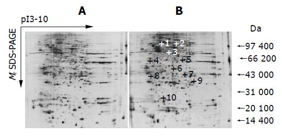
Three different independent sets of experiments were carried out to compare the pattern of cellular protein expression in untreated and H pylori-treated HepG2 cells. The two photographs shown in Figure 1 depict the pattern of cellular proteins obtained after 2-DE of total cellular extracts from untreated and H pylori-treated HepG2 cells. For untreated HepG2 cells, a total of 988±94 spots were detected; for H pylori-treated HepG2 cells, a total of 996±68 spots were detected. Most of the spots were centralized in the area of pI 4-8 and Mr 31000-97000 Da. A total of 861 spots were matched between the 2-DE maps of H pylori-treated HepG2 cells and untreated HepG2 cells. This matching analysis revealed that 10 protein spots showed higher expression in the treated cells (P<0.05). The spots that clearly overexpressed in the treated cells were marked with a cross at the corresponding site in Figure 1.

Figure 1
Figure 1 2-DE maps of untreated (A) and H pylori-treated HepG2 (B) cell proteins. The proteins from cells were extracted and separated on a pH 3-10 IPG strip, followed by a 12.5% SDS-polyacrylamide gel and silver staining. Spots that were found to be overexpressed have been annotated.
MALDI-TOF-MS analysis and protein identification

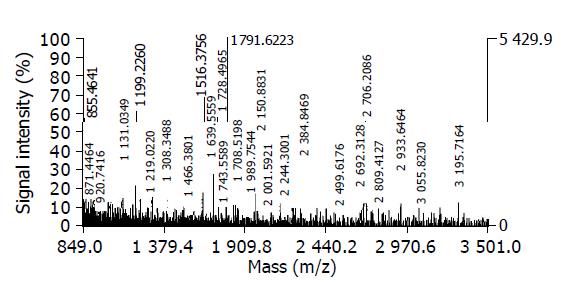
These 10 differential protein spots were excised from the stained gels and digested in-gel with trypsin. The PMF was obtained by MALDI-TOF-MS. A representative PMF map of the spot 10 was showed in Figure 2. The monoisotopic peptide masses obtained from mass spectra were used to search the SWISS-PROT and TrEMBL database with PeptIdent software. The resulting protein was determined by comprehensively considering the corresponding experimental pI, Mr, the number of matched peptides, and the sequence coverage (Tables 1 and 2). Seven proteins were successfully identified. The data on all identified protein spots were described in Table 2; these proteins were involved in transcription regulation, signal transduction, metabolism and so on.

Table 1 Matching of protein spot-10 peptide mass fingerprint data with protein Q96AX2 in database.
User massMatching massMass (Da)#MCModificationPositionPeptide
976.901977.36380.462811xCys_CAM215-222RSSCCSFM
990.94991.37950.439511xCys_PAM215-222RSSCCSFM
999.094999.53320.4392042-49TCFLIQFK
1006.9071007.37440.467411xCys_PAM,1xMSO215-222RSSCCSFM
1167.0211166.5295-0.491402xCys_CAM,1xGERA,1xMSO216-222SSCCSFM
1266.0171265.6091-0.407811xCys_CAM,1xGERA,1xMSO215-222RSSCCSFM
1308.2781308.7420.46402xGERA216-222SSCCSFM
1345.5591345.7070.1481151-162VIRSEDGETLAR
1464.5171464.84310.326112xGERA215-222RSSCCSFM
1466.3621466.81120.449202xCys_PAM,2xGERA,1xMSO216-222SSCCSFM
1516.3781516.75430.37630122-133AWLTEIHEYAQR
1684.6651684.82490.159911-17TGTPGAVATRD GEAPER
2479.0132479.22070.207711xCys_CAM18-41SPPCSPSYDLTGK VMLLGDTGVGK
Table 2 Proteins identified by MS.
SpotSWISS-PROT Accession No.Peptides MatchedpIMr (Da)Sequence coverage (%)Protein identified
1P0555619/1065.318624231.2Chain 1: integrin β-1
3P172527/286.617676439.9Protein kinase C, alpha type
6P487426/347.164448239.6LIM/homeobox protein Lhx1
7P2004210/925.603838830.9eIF-2-beta
8P46734-36/377.083994035.0MAP kinase kinase 3
9P480598/448.433725136.3PINCH protein
10Q96AX213/865.962468436.5Ras-related protein Rab-37
Figure 2
Figure 2 Peptide mass fingerprinting of the spot 10. The spot was in-gel digested with trypsin. After desalting, the peptide mixture was analyzed by MALDI-TOF-MS.
DISCUSSION

The detection that Helicobacter spp. could infect biliary tract and liver of humans has been reported by several studies in different settings. Fox et al[6], have shown the presence of Helicobacter spp. in the bile of Chileans with chronic cholecystitis. Avenaud and coworkers[3] have demonstrated by PCR the presence of genomic sequences of Helicobacter spp. in the liver of eight patients with hepatocellular carcinoma (HCC) without primary diagnosis of cirrhosis, and a further analysis by sequencing revealed that these species were H pylori and H felis. Similarly, Ponzetto[12] has reported the presence of cagA gene sequences obtained from the liver tissue of cirrhotic patients with HCC. Furthermore, Nilsson et al[2], have identified H pylori and Helicobacter spp. in human liver samples from patients suffering from PSC and PBC and recently in liver samples of patients with cholangiocarcinomas or HCC[5]. Of great interest is the fact that the gene sequence obtained from positive PCR of Helicobacter spp.-specific 16S rRNA was usually most analogous to H pylori. Helicobacter spp. has been successfully cultured from the liver of a patient with Wilson’s disease by Queiroz and Santos[23]; the isolate is closely related to H pylori by biochemical and 16S rRNA analysis. Lately, Wang et al[24], followed-up C57BL/6 and Balb/cA mice infected by three different H pylori isolates. They found that one C57BL/6 mouse infected with H pylori strain 119p developed HCC after 23 mo. Taking the results together, we may conclude that, as in animals, Helicobacter species especially H pylori may colonize the liver of humans, which could have some sort of importance in the pathogenesis and in the development of some liver diseases.

The comparative proteomic study was performed between the untreated and H pylori-treated HepG2. Ten differential protein spots were selected to perform in-gel trypsin digestion and MALDI-TOF-MS-based PMF analysis. Seven of these proteins were successfully identified by MALDI-TOF-MS. Some of the identified proteins are the oncogene-encoded products and others are the regulatory proteins of transcription and signal transduction.

The integrins are a family of cell surface glycoproteins which mediated interactions of cells with the extracellular matrix and cells with cells. These integrin-mediated interactions influence many aspects of cell behavior including cell survival, cell adhesion, cell migration, as well as cell proliferation and differentiation. All integrins are heterodimers composed of two subunits, α and β. Integrin β-1 has been found to play an essential role in invasion and metastasis of HCC cells[25]. Liu et al, found that integrin β-1 gene was upregulated in HCC using cDNA microarray technology, which was consistent with RT-PCR and Northern blot analysis of integrin β-1 gene. Human hepatoma cell lines HepG2, Huh7, and HLE stably transfected with full-length integrin β-1 had overexpression of integrin β-1 and were protected from apoptosis induced by chemotherapeutic reagents via a mitogen-activated protein kinase (MAPK)-dependent pathway[26].

Protein kinase C (PKC) is a family of at least 11 structurally related phospholipid-dependent serine/threonine kinases and it plays crucial roles in transducing signals that regulate diverse cellular functions, including proliferation, differentiation, and apoptosis. The PKC family is divided into three subgroups based on differences in their structures and co-factor requirements. PKC-α is a member of the group A or classical PKCs (cPKCs). Review of the literatures has shown that the PKC signal transduction pathway plays an important role in the development of many diseases and, in particular, cancer[27]. PKC-α is an important tumor-promoting factor; as potential target for anticancer therapy, PKC-α is currently being studied[28].

Homeobox genes play fundamental roles in development. They can be subdivided into several subfamilies, one of which is the LIM-homeobox subfamily. The characteristic features of LIM-homeodomain proteins are double zinc finger motifs, called LIM domains, that are located N-terminally of the homeodomain. These proteins mediate protein-protein interactions and are of fundamental importance for cell differentiation, cytoskeletal remodeling and transcriptional regulation[29].

Eukaryotic translation initiation factor 2 (eIF-2) is a multimeric protein consisting of three dissimilar subunits termed α, β, and γ, which functions as a central switch in the initiation of protein synthesis. In its GTP-bound state it delivers the methionyl initiator tRNA (Met-tRNA(i)) to the small ribosomal subunit and releases it upon GTP hydrolysis following the recognition of the initiation codon[30]. PINCH protein is an effector of integrin and growth factor signaling, coupling surface receptors to downstream signaling molecules involved in the regulation of cell survival, cell proliferation and cell differentiation[31].

MAPK pathway is an important signal transduction pathway that transduces extracellular signals into intracellular responses and has been implicated in a wide array of physiological processes such as proliferation, differentiation, development, apoptosis, stress and inflammatory responses. The MAPK kinases (MAPKKs) are up-stream dual-specificity protein kinases which phosphorylate and activate MAPK on threonine and tyrosine residues[32].

Ras-related protein Rab-37 belongs to the small GTPase superfamily, Rab family. GTPases regulate a myriad of cellular functions including signal transduction, cytoskeletal organization and membrane trafficking. Rab family members are also widely involved in human tumorigenesis by overexpression[33,34].

The upregulation of these proteins suggests that the capacity of the tumor invasion is stronger. However, the exact mechanisms through which H pylori lead to the change of these proteins await further study.

Advances in 2D electrophoresis, MS, and bioinformatics now make possible the large-scale examination of protein expression patterns. These improved methods are used to identify proteins found to be upregulated in expression level in H pylori-treated HepG2 cells. The above findings could be valuable for further elucidating the etiological role of Helicobacter spp. infection in a variety of liver disorders.

Footnotes

Science Editor Li WZ Language Editor Elsevier HK

References
1.  Solnick JV, Schauer DB. Emergence of diverse Helicobacter species in the pathogenesis of gastric and enterohepatic diseases. Clin Microbiol Rev. 2001;14:59-97.  [PubMed]  [DOI]  [Cited in This Article: ]  [Cited by in Crossref: 273]  [Cited by in F6Publishing: 255]  [Article Influence: 11.1]  [Reference Citation Analysis (0)]
2.  Nilsson HO, Taneera J, Castedal M, Glatz E, Olsson R, Wadström T. Identification of Helicobacter pylori and other Helicobacter species by PCR, hybridization, and partial DNA sequencing in human liver samples from patients with primary sclerosing cholangitis or primary biliary cirrhosis. J Clin Microbiol. 2000;38:1072-1076.  [PubMed]  [DOI]  [Cited in This Article: ]
3.  Avenaud P, Marais A, Monteiro L, Le Bail B, Bioulac Sage P, Balabaud C, Mégraud F. Detection of Helicobacter species in the liver of patients with and without primary liver carcinoma. Cancer. 2000;89:1431-1439.  [PubMed]  [DOI]  [Cited in This Article: ]  [Cited by in F6Publishing: 10]  [Reference Citation Analysis (0)]
4.  Myung SJ, Kim MH, Shim KN, Kim YS, Kim EO, Kim HJ, Park ET, Yoo KS, Lim BC, Seo DW. Detection of Helicobacter pylori DNA in human biliary tree and its association with hepatolithiasis. Dig Dis Sci. 2000;45:1405-1412.  [PubMed]  [DOI]  [Cited in This Article: ]  [Cited by in Crossref: 43]  [Cited by in F6Publishing: 48]  [Article Influence: 2.0]  [Reference Citation Analysis (0)]
5.  Nilsson HO, Mulchandani R, Tranberg KG, Stenram U, Wadström T. Helicobacter species identified in liver from patients with cholangiocarcinoma and hepatocellular carcinoma. Gastroenterology. 2001;120:323-324.  [PubMed]  [DOI]  [Cited in This Article: ]  [Cited by in Crossref: 81]  [Cited by in F6Publishing: 85]  [Article Influence: 3.7]  [Reference Citation Analysis (0)]
6.  Fox JG, Dewhirst FE, Shen Z, Feng Y, Taylor NS, Paster BJ, Ericson RL, Lau CN, Correa P, Araya JC. Hepatic Helicobacter species identified in bile and gallbladder tissue from Chileans with chronic cholecystitis. Gastroenterology. 1998;114:755-763.  [PubMed]  [DOI]  [Cited in This Article: ]  [Cited by in Crossref: 355]  [Cited by in F6Publishing: 334]  [Article Influence: 12.8]  [Reference Citation Analysis (0)]
7.  Roe IH, Kim JT, Lee HS, Lee JH. Detection of Helicobacter DNA in bile from bile duct diseases. J Korean Med Sci. 1999;14:182-186.  [PubMed]  [DOI]  [Cited in This Article: ]  [Cited by in Crossref: 26]  [Cited by in F6Publishing: 27]  [Article Influence: 1.1]  [Reference Citation Analysis (0)]
8.  Ito K, Nakamura M, Toda G, Negishi M, Torii A, Ohno T. Potential role of Helicobacter pylori in hepatocarcinogenesis. Int J Mol Med. 2004;13:221-227.  [PubMed]  [DOI]  [Cited in This Article: ]
9.  Silva CP, Pereira-Lima JC, Oliveira AG, Guerra JB, Marques DL, Sarmanho L, Cabral MM, Queiroz DM. Association of the presence of Helicobacter in gallbladder tissue with cholelithiasis and cholecystitis. J Clin Microbiol. 2003;41:5615-5618.  [PubMed]  [DOI]  [Cited in This Article: ]  [Cited by in Crossref: 50]  [Cited by in F6Publishing: 62]  [Article Influence: 3.1]  [Reference Citation Analysis (0)]
10.  Nilsson HO, Castedal M, Olsson R, Wadström T. Detection of Helicobacter in the liver of patients with chronic cholestatic liver diseases. J Physiol Pharmacol. 1999;50:875-882.  [PubMed]  [DOI]  [Cited in This Article: ]
11.  Chen W, Li D, Cannan RJ, Stubbs RS. Common presence of Helicobacter DNA in the gallbladder of patients with gallstone diseases and controls. Dig Liver Dis. 2003;35:237-243.  [PubMed]  [DOI]  [Cited in This Article: ]  [Cited by in Crossref: 36]  [Cited by in F6Publishing: 42]  [Article Influence: 2.0]  [Reference Citation Analysis (0)]
12.  Ponzetto A, Pellicano R, Leone N, Cutufia MA, Turrini F, Grigioni WF, D'Errico A, Mortimer P, Rizzetto M, Silengo L. Helicobacter infection and cirrhosis in hepatitis C virus carriage: is it an innocent bystander or a troublemaker? Med Hypotheses. 2000;54:275-277.  [PubMed]  [DOI]  [Cited in This Article: ]  [Cited by in Crossref: 59]  [Cited by in F6Publishing: 60]  [Article Influence: 2.5]  [Reference Citation Analysis (0)]
13.  Fan XG, Peng XN, Huang Y, Yakoob J, Wang ZM, Chen YP. Helicobacter species ribosomal DNA recovered from the liver tissue of chinese patients with primary hepatocellular carcinoma. Clin Infect Dis. 2002;35:1555-1557.  [PubMed]  [DOI]  [Cited in This Article: ]  [Cited by in Crossref: 22]  [Cited by in F6Publishing: 28]  [Article Influence: 1.3]  [Reference Citation Analysis (0)]
14.  Peng XN, Fan XG, Huang Y, Wang ZM, Chen YP. The study on relationship between Helicobacter infection and primary liver carcinoma. Shijie Huaren Xiaohua Zazhi. 2002;10:902-906.  [PubMed]  [DOI]  [Cited in This Article: ]
15.  Huang Y, Fan XG, Chen YP, Li N, Tang LJ. Detection of Helicobacter species 16S rRNA gene in paraffin-embedded hepatocellular carcinoma tissues. Shijie Huaren Xiaohua Zazhi. 2002;10:877-882.  [PubMed]  [DOI]  [Cited in This Article: ]
16.  Fagoonee S, Pellicano R, Rizzetto M, Ponzetto A. The journey from hepatitis to hepatocellular carcinoma. Bridging role of Helicobacter species. Panminerva Med. 2001;43:279-282.  [PubMed]  [DOI]  [Cited in This Article: ]
17.  Wilkins MR, Sanchez JC, Gooley AA, Appel RD, Humphery-Smith I, Hochstrasser DF, Williams KL. Progress with proteome projects: why all proteins expressed by a genome should be identified and how to do it. Biotechnol Genet Eng Rev. 1996;13:19-50.  [PubMed]  [DOI]  [Cited in This Article: ]  [Cited by in Crossref: 775]  [Cited by in F6Publishing: 640]  [Article Influence: 22.9]  [Reference Citation Analysis (0)]
18.  Pandey A, Mann M. Proteomics to study genes and genomes. Nature. 2000;405:837-846.  [PubMed]  [DOI]  [Cited in This Article: ]  [Cited by in Crossref: 1615]  [Cited by in F6Publishing: 1324]  [Article Influence: 55.2]  [Reference Citation Analysis (0)]
19.  Fan XG, Chua A, Li TG, Zeng QS. Survival of Helicobacter pylori in milk and tap water. J Gastroenterol Hepatol. 1998;13:1096-1098.  [PubMed]  [DOI]  [Cited in This Article: ]  [Cited by in Crossref: 41]  [Cited by in F6Publishing: 43]  [Article Influence: 1.7]  [Reference Citation Analysis (0)]
20.  Görg A, Obermaier C, Boguth G, Harder A, Scheibe B, Wildgruber R, Weiss W. The current state of two-dimensional electrophoresis with immobilized pH gradients. Electrophoresis. 2000;21:1037-1053.  [PubMed]  [DOI]  [Cited in This Article: ]  [Cited by in F6Publishing: 9]  [Reference Citation Analysis (0)]
21.  Corbett JM, Dunn MJ, Posch A, Görg A. Positional reproducibility of protein spots in two-dimensional polyacrylamide gel electrophoresis using immobilised pH gradient isoelectric focusing in the first dimension: an interlaboratory comparison. Electrophoresis. 1994;15:1205-1211.  [PubMed]  [DOI]  [Cited in This Article: ]  [Cited by in Crossref: 118]  [Cited by in F6Publishing: 122]  [Article Influence: 4.1]  [Reference Citation Analysis (0)]
22.  Chen P, Xie JY, Liang SP. Identification of Protein Spots in Silver-stained Two-dimensional Gels by MALDI-TOF Mass Peptide Map Analysis. ShengWu HuaXue Yu ShengWu WuLi XueBao (Shanghai). 2000;32:387-391.  [PubMed]  [DOI]  [Cited in This Article: ]
23.  de Magalhães Queiroz DM, Santos A. Isolation of a Helicobacter strain from the human liver. Gastroenterology. 2001;121:1023-1024.  [PubMed]  [DOI]  [Cited in This Article: ]  [Cited by in Crossref: 39]  [Cited by in F6Publishing: 49]  [Article Influence: 2.1]  [Reference Citation Analysis (0)]
24.  Wang X, Willén R, Svensson M, Ljungh A, Wadström T. Two-year follow-up of Helicobacter pylori infection in C57BL/6 and Balb/cA mice. APMIS. 2003;111:514-522.  [PubMed]  [DOI]  [Cited in This Article: ]  [Cited by in Crossref: 30]  [Cited by in F6Publishing: 39]  [Article Influence: 1.9]  [Reference Citation Analysis (0)]
25.  Masumoto A, Arao S, Otsuki M. Role of beta1 integrins in adhesion and invasion of hepatocellular carcinoma cells. Hepatology. 1999;29:68-74.  [PubMed]  [DOI]  [Cited in This Article: ]  [Cited by in Crossref: 60]  [Cited by in F6Publishing: 60]  [Article Influence: 2.4]  [Reference Citation Analysis (0)]
26.  Zhang H, Ozaki I, Mizuta T, Matsuhashi S, Yoshimura T, Hisatomi A, Tadano J, Sakai T, Yamamoto K. Beta 1-integrin protects hepatoma cells from chemotherapy induced apoptosis via a mitogen-activated protein kinase dependent pathway. Cancer. 2002;95:896-906.  [PubMed]  [DOI]  [Cited in This Article: ]  [Cited by in Crossref: 53]  [Cited by in F6Publishing: 57]  [Article Influence: 2.6]  [Reference Citation Analysis (0)]
27.  Rybczyńska M, Ksiazek K, Kaczmarek J. The role of PKC isoforms in tumorigenicity and apoptotic cell death. Postepy Hig Med Dosw. 2000;54:777-796.  [PubMed]  [DOI]  [Cited in This Article: ]
28.  Roychowdhury D, Lahn M. Antisense therapy directed to protein kinase C-alpha (Affinitak, LY900003/ISIS 3521): potential role in breast cancer. Semin Oncol. 2003;30:30-33.  [PubMed]  [DOI]  [Cited in This Article: ]  [Cited by in Crossref: 7]  [Cited by in F6Publishing: 8]  [Article Influence: 0.4]  [Reference Citation Analysis (0)]
29.  Hobert O, Westphal H. Functions of LIM-homeobox genes. Trends Genet. 2000;16:75-83.  [PubMed]  [DOI]  [Cited in This Article: ]  [Cited by in Crossref: 367]  [Cited by in F6Publishing: 358]  [Article Influence: 14.9]  [Reference Citation Analysis (0)]
30.  Sonenberg N, Dever TE. Eukaryotic translation initiation factors and regulators. Curr Opin Struct Biol. 2003;13:56-63.  [PubMed]  [DOI]  [Cited in This Article: ]  [Cited by in Crossref: 246]  [Cited by in F6Publishing: 250]  [Article Influence: 11.9]  [Reference Citation Analysis (0)]
31.  Wang-Rodriguez J, Dreilinger AD, Alsharabi GM, Rearden A. The signaling adapter protein PINCH is up-regulated in the stroma of common cancers, notably at invasive edges. Cancer. 2002;95:1387-1395.  [PubMed]  [DOI]  [Cited in This Article: ]  [Cited by in Crossref: 54]  [Cited by in F6Publishing: 58]  [Article Influence: 2.6]  [Reference Citation Analysis (0)]
32.  Johnson GL, Lapadat R. Mitogen-activated protein kinase pathways mediated by ERK, JNK, and p38 protein kinases. Science. 2002;298:1911-1912.  [PubMed]  [DOI]  [Cited in This Article: ]  [Cited by in Crossref: 3062]  [Cited by in F6Publishing: 3232]  [Article Influence: 146.9]  [Reference Citation Analysis (0)]
33.  Pfeffer SR. Rab GTPases: specifying and deciphering organelle identity and function. Trends Cell Biol. 2001;11:487-491.  [PubMed]  [DOI]  [Cited in This Article: ]  [Cited by in Crossref: 422]  [Cited by in F6Publishing: 438]  [Article Influence: 19.0]  [Reference Citation Analysis (0)]
34.  He H, Dai F, Yu L, She X, Zhao Y, Jiang J, Chen X, Zhao S. Identification and characterization of nine novel human small GTPases showing variable expressions in liver cancer tissues. Gene Expr. 2002;10:231-242.  [PubMed]  [DOI]  [Cited in This Article: ]