Published online Jun 14, 2005. doi: 10.3748/wjg.v11.i22.3446
Revised: June 20, 2004
Accepted: July 22, 2004
Published online: June 14, 2005
AIM: To evaluate the possible value of FasL in gastric cancer gene therapy by investigating the effects of FasL expression on human gastric cancer cell line.
METHODS: An adenoviral vector encoding the full-length human FasL cDNA was constructed and used to infect a human gastric cancer (SGC-7901) cell line. FasL expression was confirmed by X-gal staining, flow cytometric analysis and RT-PCR. The effect of FasL on cell proliferation was determined by clonogenic assay, cytotoxicity was detected by MTT assay, and cell viability was measured by trypan blue exclusion. The therapeutic efficiency of Ad-FasL in vivo was investigated with a xenograft tumor model in nude mice.
RESULTS: SGC-7901 cells infected with Ad-FasL showed increased expression of FasL, resulting in significantly decreased cell growth and colony-forming activity when compared with control adenovirus-infected cells. The cytotoxicity of anti-Fas antibody (CH-11) in gastric cancer cells was stronger than that of ActD (91±8 vs 60±5, P<0.01), and the cytotoxicity of Ad-FasL was stronger than that of CH-11 (60±5 vs 50±2, P<0.05). In addition, G1-phase arrest (67.75±0.39 vs 58.03±2.16, P<0.05) and apoptosis were observed in Ad-FasL-infected SGC-7901 cells, and the growth of SGC-7901 xenografts in nude mice was retarded after intra-tumoral injection with Ad-FasL (54% vs 0%, P<0.0001).
CONCLUSION: Infection of human gastric carcinoma cells with Ad-FasL induces apoptosis, indicating that this target gene might be of potential value in gene therapy for gastric cancer.
- Citation: Zheng SY, Li DC, Zhang ZD, Zhao J, Ge JF. Adenovirus-mediated FasL gene transfer into human gastric carcinoma. World J Gastroenterol 2005; 11(22): 3446-3450
- URL: https://www.wjgnet.com/1007-9327/full/v11/i22/3446.htm
- DOI: https://dx.doi.org/10.3748/wjg.v11.i22.3446
Gastric cancer is one of the most common digestive tract cancers in China. Although an increasing number of gastric cancer patients have benefited from the development of modern tumor therapies, the prognosis of this disease is still relatively poor. Gastric cancer often resists various treatments, including immunotherapy, wherein deficient tumor-specific T-cell responses result in poor immune response.
In this context, apoptosis mediated by the Fas/FasL system is of great interest to researchers, as cytotoxic T-lymphocytes (CTLs) utilize the perforin/granzyme and Fas/FasL systems to kill cancer cells. Fas is a member of the tumor necrosis factor/nerve growth factor receptor family, whereas the Fas ligand (FasL) is a member of the tumor necrosis factor/nerve growth factor family[1-7]. The FasL is primarily expressed in active CTLs, and induces apoptosis of Fas-expressing tumor cells[2,6]. Since the Fas/FasL apoptosis pathway is a key mechanism for clearing tumor cells, researchers are currently seeking methods for triggering FasL expression in tumor cells via gene therapy, thus ‘marking’ them for Fas/FasL-mediated apoptosis[5,8]. Accordingly, we used adenoviral gene transfer to trigger high-level FasL expression in SGC-7901 (human gastric cancer) cells to investigate the possible use of FasL in gastric cancer gene therapy.
The human gastric cancer cell line SGC-7901 was obtained from the Shanghai Institute of Cell Biology at the Chinese Academy of Sciences (Shanghai, China). Cells were cultured in Medium 199 (Gibco) supplemented with 10% fetal bovine serum.
The recombinant FasL retroviral vector was constructed in our laboratory. The FasL gene expression cassette includes the CMV promoter, a full-length FasL cDNA (Jingmei Company, Shenzheng, China) and the SV40 polyA signal sequences. This cassette was inserted into the E1 region of an adenoviral genome lacking the viral E1 and E3 sequences. Briefly, the FasL cDNA was inserted into the pAdCMV shuttle plasmid (kindly provided by Dr. Daru Lu) and co-transfected with pJM17 (Microbix Biosystems Inc., Canada) into human embryonic 293 cells (provided by Dr. Lu) using the Lipofectin reagent (Gibco). The FasL expression cassette was then transferred into the adenovirus genome by homologous recombination. The control virus Ad-LacZ was constructed in the same manner. Virus proliferation, purification and titering were performed as described by He et al[9].
Gastric carcinoma cells were infected with Ad-LacZ at various multiplicities of infection (MOIs). After 48 h, cells were fixed in 40 g/L formaldehyde for 6-8 h and then treated with X-gal solution (1 mg/mL X-gal in a solution of 0.1 mol/L PBS, 1.3 mmol/L MgCl2, 3 mmol/L K3Fe(CN)6) for 2 h or overnight in a 37 °C incubator. The percentage of blue cells was then determined.
Cells were passaged at a density of 105/well. For viral infection, cells were incubated with virus suspensions (at various MOIs) and 8 μg/mL polybrene (Sigma) for 2-3 h at 37 °C, washed twice with fresh medium, and further incubated for 48 h. Then cells were passaged and incubated in medium containing 1 mg/mL G418 (Gibco) for selection of transfectants. The medium was changed every 3-4 d until anti-G418 cells appeared.
Cells were harvested, washed with PBS, and treated with FITC-conjugated FasL antibody (Jackson ImmunoResearch Lab) or control serum for 0.5-1 h at 4 °C. Cells were once again washed with PBS, and FasL protein expression was confirmed by FCM analysis (Becton Dickinson, San Jose, CA). For FasL gene expression, RT-PCR was performed with the kit according to the manufacturer’s instructions (Shanghai Shanggong Company, Shanghai, China). The primers were: forward 5’-CTGAATTCTGACTCACCA-GCTGCCATGC-3’, reverse 5’-TACTCGAGCTATTAGA-GCTTATATAAGCCG-3’.
SGC-7901 cells were infected with Ad-FasL or Ad-LacZ (100 MOI). After 24 h, the cells were seeded in 6-well plates at 500 cells/well. After being incubated for 2-3 wk, cells were stained with 0.1% crystal violet and counted under a microscope. Colonies of more than 50 cells were counted for all clonogenic assays.
Cells infected with Ad-FasL or Ad-LacZ (more than 106 cells) in either suspension or adhesion were harvested and fixed in 70% ethanol for 3 h. Cells were then treated with 50 μg/mL RNase for 1 h at 37 °C, and stained with 100 μg/mL PI for 20-30 min prior to FCM analysis.
SGC-7901 cells were treated with 100 ng/mL anti-Fas antibody (CH-11; PharMingen), 50 ng/mL actinomycin D (ActD; Sigma), and infected with Ad-FasL (100 MOI) or treated with anti-Fas antibody combined with Ad-FasL infection. Cytotoxicity was determined by MTT assay. About 103-4 cells/well were plated in 96-well plates and incubated overnight in 100 μL of culture medium. After 2-3 d, 20 μL of MTT solution (5 mg/mL) was added to each culture well. After incubating for 4 h at 37 °C, the MTT was removed and 200 μL of dimethyl sulfoxide (Sigma) was added and mixtures were shaken, the crystals were fully dissolved (about 10 min). The A value of each well was detected using a microculture plate reader (Huandong Cesium Electron Tube Company) with a test wavelength of 490 nm. Cell survival rate (SR) was expressed as the following equation: SR=(A in experimental group/A in control group)×100%. Results were expressed as mean±SD; the Student’s t-test was used for statistical analysis.
SGC-7901 cells (2.5-3.0×10³ cells/well) were plated in 24-well plates (3 wells for each test) and infected with various MOIs of Ad-LacZ or Ad-FasL for 24 h. Cells were harvested every 2 d, and the living cell rate (LR) was measured by trypan blue exclusion assay. LR was expressed as the following equation:
LR=number of living cells/(number of living cells+ number of dead cells)×100%. Results were expressed as mean±SD; ANOVA was used for statistical analysis with SPSS 10.0 software. P<0.05 was considered statistically significant.
BALB/c nude mice (The Shanghai Institute of Cancer Research) were subcutaneously injected with SGC-7901 cells at 1-5×10⁷ cells/mouse. When tumors grew to 0.5 cm in diameter, the mice were randomly divided into treated and control groups (n = 5). Tumor volumes were calculated by [(1/2)×(longest diameter)×(shortest diameter)] as described previously. Tumors were measured every 5 d for 6 wk. Growth curves were drawn and the percentage of tumor inhibition was calculated (treated group/control group×100%.
Anti-G418 colonies were obtained after screening for 2 wk. The selected clone containing the FasL gene was named as SGC-7901-FasL, and the control clone containing the blank vector was designated as SGC-7901-vect. There was no morphologic difference between cultures of these two cells.
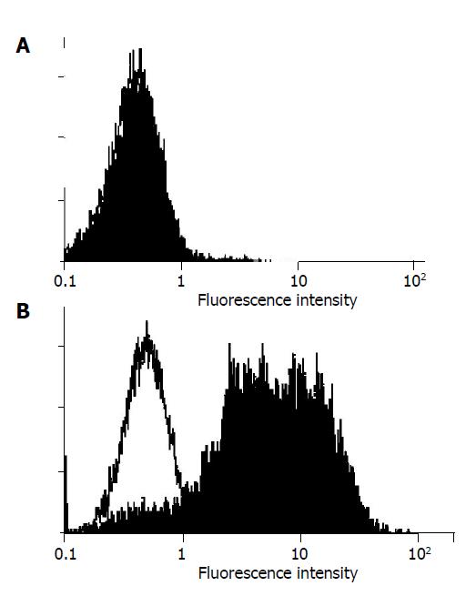
FCM analysis revealed that SGC-7901-FasL cells expressed FasL on their surface, whereas SGC-7901-vect did not (Figure 1).
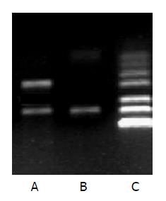
Total RNA was extracted from the test and control cells, and FasL-specific primers were used for amplification by RT-PCR. The expected 231-bp fragment was amplified from SGC-7901-FasL cells, but not from SCG-7901-vect cells (Figure 2).
Two days after infection, SGC-7901-FasL cells were smaller and became more round in shape. Over the next 4 d, the plasma membranes of these cells blebbed, the cytoplasm and nuclei condensed, and the cells ultimately lysed into membrane-bound apoptotic bodies. Cytotoxicity was determined by MTT 4 d after infection. Ad-FasL cultures showed 84% fewer viable cells than Ad-LacZ infected cultures, suggesting that Ad-FasL was cytotoxic to gastric cancer cells. Clonogenic assay showed that SGC-7901 cells infected with Ad-FasL (100 MOI) did not form colonies, whereas Ad-LacZ cultures formed numerous colonies, demonstrating that expression of FasL significantly decreased colony formation.
As shown in Table 1, Ad-FasL-infected cultures showed fewer cells in S or G2 M phase, and more cells in G1 phase, indicating that FasL could inhibit amplification of gastric tumor cells. A sub-G1 peak (apoptosis peak; Table 1) appeared 4 d after infection of Ad-FasL, indicating that the FasL gene not only induced G1 phase arrest, but also induced apoptosis of SGC-7901 cells.
To further evaluate the possibility of gastric cancer gene therapy with Ad-FasL, SGC-7901 cells were infected with Ad-FasL or treated with anti-Fas antibody, and the resulting cytotoxicity was compared with that of ActD, a RNA synthesis inhibitor known as cytotoxin. The cytotoxicity of the anti-Fas antibody (CH-11) to gastric cancer cells was stronger than that of ActD, and the cytotoxicity of Ad-FasL was stronger than that of CH-11 (Table 2).
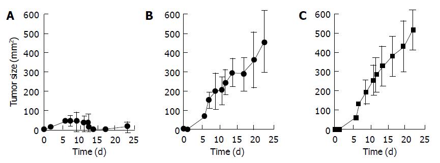
Nude mice were subcutaneously injected with SGC-7901 cells, then intratumoral injections of Ad-FasL were administered. The growth of tumor infected with Ad-FasL was inhibited by 54%, suggesting that Ad-FasL was a viable gene therapy candidate (Figure 3).
Although a growing number of tumor patients have benefited from modern oncotherapeutic methods, there is still a need to improve therapies for malignant tumors. Gene therapy is expected to join surgical, radiological and chemotherapeutic strategies in future methods of integrated oncotherapy. Pre-clinical studies have confirmed that adenovirus-mediated high level expression of carcinoma-inhibiting genes (such as p53) can inhibit tumor growth, induce apoptosis and increase tumor tissue sensitivity to radio- and chemotherapy[10-14]. Recent clinical studies in China and abroad have indicated that adenoviral gene therapy is safe and applicable[12-15]. Here, we used this vector to express FasL in cultured human gastric cancer cells. The FasL gene expression cassette includes the CMV promoter, a full-length FasL cDNA, and SV40 polyA signal sequences. A transduction efficiency test using a similarly constructed Ad-LacZ vector illustrated that the adenoviral construct possessed high transduction efficiency. FCM and RT-PCR were used to detect high-level FasL expression in target cells, confirming that the adenovirus vector effectively transfers the FasL gene into tumor cells.
Previous studies[16,17] indicated that binding between FasL and Fas induces receptor trimerization, and apoptosis of Fas-expressing cells. Current theory holds that the signaling responsible for this apoptosis occurs in one of the following three ways: between T cells and target cells, among target cells; or between T cells[18-23]. Fas expression is markedly higher in gastric cancer cells than in normal gastric mucosal cells, implying that Fas participates in the genesis of gastric carcinoma. Fas activation can induce gastric carcinoma cell apoptosis, indicating that the Fas/FasL system might be a good target for gene therapy. In this study, we attempted to induce direct apoptosis of target cells (cis-type apoptosis) by transfecting a highly efficient Ad-FasL expression vector into gastric carcinoma cells (SGC-7901). Expression of FasL inhibited the apoptosis of SGC-7901 cells up to 84%, and significantly inhibited the ability of SGC-7901 cells to form colonies. These results have not been reported in China.
FasL is thought to engage with Fas by inducing receptor trimerization, which then transfers signals to the Fas intracellular death domains (DD). Then, the Fas-associated death domain dimerizes with the DD to transfer an apoptotic signal to Caspase-8, instigating a caspase cascade leading to cell apoptosis[24-26]. In our study, FCM analysis showed that SGC-7901 cells infected with Ad-FasL quickly arrested in the G1 phase, which was subsequently followed by tumor cell apoptosis. Taken together, these in vitro results suggest that FasL gene transfer is capable of inducing gastric tumor cell apoptosis and that it may be a viable candidate for tumor gene therapy.
In our study, an in vitro cytotoxicity assay showed that Ad-FasL could significantly inhibit the growth of gastric cancer cells. The inhibition was much stronger than the cytotoxicity conferred by CH-11 treatment, indicating that the Fas/FasL system plays an important role in gastric cancer cell apoptosis[27-29]. In contrast, a SGC-7901 tumor model in nude mice showed only 54% inhibition of tumor growth in response to Ad-FasL infection. This difference between the in vivo and in vitro response rates might be caused by poor distribution of the recombinant adenovirus in the solid tumor, resulting in lack of target gene transfer to all the tumor cells. Thus, future Fas/FasL gene therapy experiments in vivo should focus on stabilizing vectors, increasing transfection efficiency, repeating administration and combining interventional therapy and gene therapy, thereby improving the therapeutic efficacy.
| 1. | Sharma K, Wang RX, Zhang LY, Yin DL, Luo XY, Solomon JC, Jiang RF, Markos K, Davidson W, Scott DW. Death the Fas way: regulation and pathophysiology of CD95 and its ligand. Pharmacol Ther. 2000;88:333-347. [RCA] [PubMed] [DOI] [Full Text] [Cited by in Crossref: 140] [Cited by in RCA: 143] [Article Influence: 5.7] [Reference Citation Analysis (0)] |
| 2. | Sapi E, Brown WD, Aschkenazi S, Lim C, Munoz A, Kacinski BM, Rutherford T, Mor G. Regulation of Fas ligand expression by estrogen in normal ovary. J Soc Gynecol Investig. 2002;9:243-250. [RCA] [PubMed] [DOI] [Full Text] [Cited by in Crossref: 22] [Cited by in RCA: 23] [Article Influence: 1.0] [Reference Citation Analysis (0)] |
| 3. | Gniadecki R. Depletion of membrane cholesterol causes ligand-independent activation of Fas and apoptosis. Biochem Biophys Res Commun. 2004;320:165-169. [RCA] [PubMed] [DOI] [Full Text] [Cited by in Crossref: 97] [Cited by in RCA: 100] [Article Influence: 4.8] [Reference Citation Analysis (0)] |
| 4. | Walczak H, Krammer PH. The CD95 (APO-1/Fas) and the TRAIL (APO-2L) apoptosis systems. Exp Cell Res. 2000;256:58-66. [RCA] [PubMed] [DOI] [Full Text] [Cited by in Crossref: 446] [Cited by in RCA: 439] [Article Influence: 17.6] [Reference Citation Analysis (0)] |
| 5. | Li-Weber M, Krammer PH. Function and regulation of the CD95 (APO-1/Fas) ligand in the immune system. Semin Immunol. 2003;15:145-157. [RCA] [PubMed] [DOI] [Full Text] [Cited by in Crossref: 86] [Cited by in RCA: 82] [Article Influence: 3.9] [Reference Citation Analysis (0)] |
| 6. | Reichmann E. The biological role of the Fas/FasL system during tumor formation and progression. Semin Cancer Biol. 2002;12:309-315. [RCA] [PubMed] [DOI] [Full Text] [Cited by in Crossref: 80] [Cited by in RCA: 79] [Article Influence: 3.4] [Reference Citation Analysis (0)] |
| 7. | Gu XH, Li QF, Wang YM. Expression of hepatocyte apoptosis and Fas/FasL, in liver tissues of patients with hepatitis D. Shijie Huaren Xiaohua Zazhi. 2000;8:35-38. |
| 8. | Aoki K, Akyürek LM, San H, Leung K, Parmacek MS, Nabel EG, Nabel GJ. Restricted expression of an adenoviral vector encoding Fas ligand (CD95L) enhances safety for cancer gene therapy. Mol Ther. 2000;1:555-565. [RCA] [PubMed] [DOI] [Full Text] [Cited by in Crossref: 42] [Cited by in RCA: 41] [Article Influence: 1.6] [Reference Citation Analysis (0)] |
| 9. | He TC, Zhou S, da Costa LT, Yu J, Kinzler KW, Vogelstein B. A simplified system for generating recombinant adenoviruses. Proc Natl Acad Sci USA. 1998;95:2509-2514. [RCA] [PubMed] [DOI] [Full Text] [Cited by in Crossref: 2861] [Cited by in RCA: 3045] [Article Influence: 112.8] [Reference Citation Analysis (0)] |
| 10. | Dunkern T, Roos W, Kaina B. Apoptosis induced by MNNG in human TK6 lymphoblastoid cells is p53 and Fas/CD95/Apo-1 related. Mutat Res. 2003;544:167-172. [RCA] [PubMed] [DOI] [Full Text] [Cited by in Crossref: 19] [Cited by in RCA: 18] [Article Influence: 0.9] [Reference Citation Analysis (0)] |
| 11. | Juang SH, Pan WY, Kuo CC, Liou JP, Hung YM, Chen LT, Hsieh HP, Chang JY. A novel bis-benzylidenecyclopentanone derivative, BPR0Y007, inducing a rapid caspase activation involving upregulation of Fas (CD95/APO-1) and wild-type p53 in human oral epidermoid carcinoma cells. Biochem Pharmacol. 2004;68:293-303. [RCA] [PubMed] [DOI] [Full Text] [Cited by in Crossref: 11] [Cited by in RCA: 10] [Article Influence: 0.5] [Reference Citation Analysis (0)] |
| 12. | Rubinchik S, Wang D, Yu H, Fan F, Luo M, Norris JS, Dong JY. A complex adenovirus vector that delivers FASL-GFP with combined prostate-specific and tetracycline-regulated expression. Mol Ther. 2001;4:416-426. [RCA] [PubMed] [DOI] [Full Text] [Cited by in Crossref: 41] [Cited by in RCA: 39] [Article Influence: 1.6] [Reference Citation Analysis (0)] |
| 13. | Xu GW, Sun ZT, Forrester K, Wang XW, Coursen J, Harris CC. Tissue-specific growth suppression and chemosensitivity promotion in human hepatocellular carcinoma cells by retroviral-mediated transfer of the wild-type p53 gene. Hepatology. 1996;24:1264-1268. [RCA] [PubMed] [DOI] [Full Text] [Cited by in Crossref: 50] [Cited by in RCA: 51] [Article Influence: 1.8] [Reference Citation Analysis (0)] |
| 14. | Habib NA, Hodgson HJ, Lemoine N, Pignatelli M. A phase I/II study of hepatic artery infusion with wtp53-CMV-Ad in metastatic malignant liver tumours. Hum Gene Ther. 1999;10:2019-2034. [RCA] [PubMed] [DOI] [Full Text] [Cited by in Crossref: 52] [Cited by in RCA: 46] [Article Influence: 1.8] [Reference Citation Analysis (0)] |
| 15. | Schuler M, Rochlitz C, Horowitz JA, Schlegel J, Perruchoud AP, Kommoss F, Bolliger CT, Kauczor HU, Dalquen P, Fritz MA. A phase I study of adenovirus-mediated wild-type p53 gene transfer in patients with advanced non-small cell lung cancer. Hum Gene Ther. 1998;9:2075-2082. [RCA] [PubMed] [DOI] [Full Text] [Cited by in Crossref: 119] [Cited by in RCA: 104] [Article Influence: 3.9] [Reference Citation Analysis (0)] |
| 16. | Miura Y, Thoburn CJ, Bright EC, Hess AD. Cytolytic effector mechanisms and gene expression in autologous graft-versus-host disease: distinct roles of perforin and Fas ligand. Biol Blood Marrow Transplant. 2004;10:156-170. [RCA] [PubMed] [DOI] [Full Text] [Cited by in Crossref: 12] [Cited by in RCA: 10] [Article Influence: 0.5] [Reference Citation Analysis (0)] |
| 17. | Webb SD, Sherratt JA, Fish RG. Cells behaving badly: a theoretical model for the Fas/FasL system in tumour immunology. Math Biosci. 2002;179:113-129. [RCA] [PubMed] [DOI] [Full Text] [Cited by in Crossref: 27] [Cited by in RCA: 28] [Article Influence: 1.2] [Reference Citation Analysis (0)] |
| 18. | Wang LS, Liu HJ, Zhang JH, Wu CT. Purging effect of dibutyl phthalate on leukemia cells involves fas independent activation of caspase-3/CPP32 protease. Cancer Lett. 2002;186:177-182. [RCA] [PubMed] [DOI] [Full Text] [Cited by in Crossref: 10] [Cited by in RCA: 10] [Article Influence: 0.4] [Reference Citation Analysis (0)] |
| 19. | Chun YJ, Park S, Yang SA. Activation of Fas receptor modulates cytochrome P450 3A4 expression in human colon carcinoma cells. Toxicol Lett. 2003;146:75-81. [RCA] [PubMed] [DOI] [Full Text] [Cited by in Crossref: 8] [Cited by in RCA: 8] [Article Influence: 0.4] [Reference Citation Analysis (0)] |
| 20. | Modiano JF, Sun J, Lang J, Vacano G, Patterson D, Chan D, Franzusoff A, Gianani R, Meech SJ, Duke R. Fas ligand-dependent suppression of autoimmunity via recruitment and subsequent termination of activated T cells. Clin Immunol. 2004;112:54-65. [RCA] [PubMed] [DOI] [Full Text] [Cited by in Crossref: 10] [Cited by in RCA: 10] [Article Influence: 0.5] [Reference Citation Analysis (0)] |
| 21. | Granville DJ, Jiang H, McManus BM, Hunt DW. Fas ligand and TRAIL augment the effect of photodynamic therapy on the induction of apoptosis in JURKAT cells. Int Immunopharmacol. 2001;1:1831-1840. [RCA] [PubMed] [DOI] [Full Text] [Cited by in Crossref: 20] [Cited by in RCA: 21] [Article Influence: 0.9] [Reference Citation Analysis (0)] |
| 22. | Webb SD, Sherratt JA. A perturbation problem arising from the modelling of soluble Fas ligand in tumour immunology. Mathem Comp Modell. 2003;37:323-331. [RCA] [DOI] [Full Text] [Cited by in Crossref: 3] [Cited by in RCA: 2] [Article Influence: 0.1] [Reference Citation Analysis (0)] |
| 23. | Kase H, Aoki Y, Tanaka K. Fas ligand expression in cervical adenocarcinoma: relevance to lymph node metastasis and tumor progression. Gynecol Oncol. 2003;90:70-74. [RCA] [PubMed] [DOI] [Full Text] [Cited by in Crossref: 26] [Cited by in RCA: 26] [Article Influence: 1.2] [Reference Citation Analysis (0)] |
| 24. | Wiener Z, Ontsouka EC, Jakob S, Torgler R, Falus A, Mueller C, Brunner T. Synergistic induction of the Fas (CD95) ligand promoter by Max and NFkappaB in human non-small lung cancer cells. Exp Cell Res. 2004;299:227-235. [RCA] [PubMed] [DOI] [Full Text] [Cited by in Crossref: 11] [Cited by in RCA: 14] [Article Influence: 0.7] [Reference Citation Analysis (0)] |
| 25. | Chatterjee D, Schmitz I, Krueger A, Yeung K, Kirchhoff S, Krammer PH, Peter ME, Wyche JH, Pantazis P. Induction of apoptosis in 9-nitrocamptothecin-treated DU145 human prostate carcinoma cells correlates with de novo synthesis of CD95 and CD95 ligand and down-regulation of c-FLIP(short). Cancer Res. 2001;61:7148-7154. [PubMed] |
| 26. | Liu HF, Liu WW, Fang DC. Effect of combined anti Fas mAb and IFN-γ on the induction of apoptosis in human gastric carcinoma cell line SGC-7901. Shijie Huaren Xiaohua Zazhi. 2000;8:1361-1364. |
| 27. | Kim KB, Choi YH, Kim IK, Chung CW, Kim BJ, Park YM, Jung YK. Potentiation of Fas- and TRAIL-mediated apoptosis by IFN-gamma in A549 lung epithelial cells: enhancement of caspase-8 expression through IFN-response element. Cytokine. 2002;20:283-288. [RCA] [PubMed] [DOI] [Full Text] [Cited by in Crossref: 35] [Cited by in RCA: 35] [Article Influence: 1.5] [Reference Citation Analysis (0)] |
| 28. | González-Cuadrado S, López-Armada MJ, Gómez-Guerrero C, Subirá D, Garcia-Sahuquillo A, Ortiz-Gonzalez A, Neilson EG, Egido J, Ortiz A. Anti-Fas antibodies induce cytolysis and apoptosis in cultured human mesangial cells. Kidney Int. 1996;49:1064-1070. [RCA] [PubMed] [DOI] [Full Text] [Cited by in Crossref: 46] [Cited by in RCA: 43] [Article Influence: 1.5] [Reference Citation Analysis (0)] |
| 29. | Schmitz I, Krueger A, Baumann S, Kirchhoff S, Krammer PH. Specificity of anti-human CD95 (APO-1/Fas) antibodies. Biochem Biophys Res Commun. 2002;297:459-462. [RCA] [PubMed] [DOI] [Full Text] [Cited by in Crossref: 6] [Cited by in RCA: 5] [Article Influence: 0.2] [Reference Citation Analysis (0)] |


