Published online Apr 21, 2005. doi: 10.3748/wjg.v11.i15.2357
Revised: September 8, 2004
Accepted: October 8, 2004
Published online: April 21, 2005
Intracardiac manifestation of hepatocellular carcinoma (HCC) is a rare condition and an uncommon finding even at autopsy. Pulmonary tumor embolism as a presenting feature of HCC has been published only twice previously. In our case report, a 63-year-old man presented with high fever and six episodes of recurrent pneumonias during the last half year. Echocardiography was performed, a solid mass was found in the right atrium. Transesophageal echocardiography proved a tumor mass in the inferior vena cava (IVC) extending into the right atrium, abdominal ultrasound revealed tumor mass in the IVC and a solid tumor in the liver. Combined liver and heart surgery was attempted in order to remove the tumor mass from both the liver and the right atrium. Acute cor pulmonale occurred during tumor removal from the right atrium and the patient expired. In addition to local factors the possibility of embolization should arise in the background of recurrent pneumonia. Occult carcinoma must be included in pos-sible causes of recurrent pulmonary embolism. Searching for primary malignancy should include HCC as frequent cause of hypercoagulability. In case of HCC, echocar-diography is suggested because of the possibility of expansion in IVC or right atrium and tumor-embolization.
- Citation: Papp E, Keszthelyi Z, Kalmar NK, Papp L, Weninger C, Tornoczky T, Kalman E, Toth K, Habon T. Pulmonary embolization as primary manifestation of hepatocellular carcinoma with intracardiac penetration: A case report. World J Gastroenterol 2005; 11(15): 2357-2359
- URL: https://www.wjgnet.com/1007-9327/full/v11/i15/2357.htm
- DOI: https://dx.doi.org/10.3748/wjg.v11.i15.2357
Intracardiac manifestation of hepatocellular carcinoma (HCC) is a rare condition, uncommon finding even at autopsy. Pulmonary tumor embolism as a presenting feature of HCC has been published only twice previously[1,2]. In our case recurrent pneumonias and dyspnea were the only symptoms distantly related to HCC.
A 63-year-old man presented with high fever, six episodes of recurrent pneumonias during the last half year. His medical history showed pulmonary tuberculosis (30 years ago) and hypertension. There was no hepatitis or any other liver dysfunction in his case history. Only the enlarged liver was a pathologic finding at physical examination.
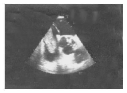
Two-dimensional transthoracic echocardiography was performed searching for possible infective endocarditis in the background of recurrent fevers. A solid mass was found in the right atrium causing severe inflow obstruction. Transesophageal echocardiography proved a tumor mass in the inferior vena cava (IVC) extending into the right atrium (Figure 1).
Elevated red blood cell sedimentation rate, normal hema-tocrit, normal transaminase levels and extremely high alfa-fetoprotein (AFP) level (18184 ng/mL, normal range: <8 ng/mL) were seen in the blood chemistry. No labor-atory signs of liver disease were present. Serological examin-ations disclosed hepatitis B or C viral infection.
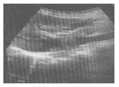
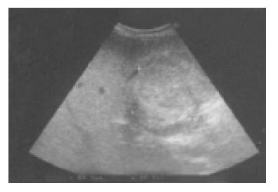
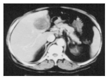
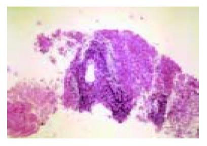
Abdominal ultrasound revealed tumor mass in IVC and a solid tumor in the fourth segment of the right lobe of the liver (Figures 2 and 3). Abdominal computerized tomography (CT) scan proved this finding (Figure 4). Fine needle bio-psy was performed, which showed small round cell HCC (Figure 5). Combined liver and heart surgery was attempted in order to remove the tumor mass from both the liver and the right atrium. Off pump heart surgery was chosen to avoid an adverse immunocompromised state known to be related to extracorporeal bypass. Acute cor pulmonale occu-rred during tumor removal from the right atrium and the patient expired. Autopsy revealed fresh pulmonary infarction in the left lung with thrombi in the left main pulmonary artery supplying the infarcted part. Massive embolus was seen in the central part of the right pulmonary artery as a cause of acute cor pulmonale. A tumor mass with a diameter of -7 cm was detected in the right lobe of the liver with no signs of cirrhosis. Histologic examinations proved tumor cells in the thrombus from the IVC (Figure 6), both small round cells and conventional HCC cells in the liver tumor mass with AFP and CD99 positivity (Figure 7).
Cancer patients are prone to develop both thrombotic and tumor pulmonary embolism. Although metastatic spread of malignancies to lungs is common, pulmonary tumor embolism is an unusual phenomenon. It remains undetected often and many cases are not recognized until postmortem examination[3].
This case is the third reported one of tumor emboli as first sign of HCC and the only reported case in which neither the signs of pulmonary embolism nor any of the typical manifestations of HCC but only recurrent pneumonia were present. Autopsy proved tumor emboli in the background of pneumonia.
HCC is one of the most common cancers worldwide but it has a low prevalence in the Western world. More than 70% of patients with HCC have cirrhosis, and more than 80% have elevated alkaline phosphatase or aspartateaminotransferase serum levels. Epidemiological studies have established that hepatitis B and C viral infections are the main etiologic factors. Patients typically present with abdominal pain and weight loss. None of these were present in the case reported.
Although invasion of the IVC and the right atrium by HCC occurs in less than 2% of the patients with HCC[4], only few cases were reported[4-6]. Pulmonary manifestations vary, the “cannonball” pattern being the most common one[7-9]. Hypercoagulability in malignancy is well known and liver cancer is strongly associated with venous thromboembolism[10]. It is surprising that only two cases with pulmonary embolism have previously been reported.
Intraatrial manifestation of the HCC is a life-threatening condition. The major causes of death are either sudden pulmonary embolism of the thrombus or acute obstruction of the tricuspid valve or both. Resection can provide relatively good long-term survival in this particular clinical situation but not more than 2 years[11]. The surgical approach to IVC and intraatrial masses is difficult. These patients usually die within a short period because of pulmonary embolism, heart failure or cancer progression. The only treatment option is hepatic resection with removal of the tumor thrombus. Most commonly, deep hypothermia and circulatory arrest have been used[12,13]. Georgen et al[14], reported a successful removal of HCC from the right atrium without extracorporeal bypass. Optimal decision making in rare cases like this is a complicated task based on intuition rather than scanty information provided by evidence-based medicine.
In summary, in addition to local factors the possibility of embolization should arise in the background of recurrent pneumonia. Occult carcinoma must be included in possible causes of recurrent pulmonary embolism. Searching for primary malignancy should include HCC as the frequent cause of hypercoagulability. Regarding the literature review referring to HCC cases, echocardiography is suggested because of the possibility of expansion in IVC or right atrium and tumor-embolization[15]. This case is reported because it describes a rapid clinical progression where the recurrent pneumonia was the first manifestation of a previously undiagnosed neoplastic disease.
Science Editor Guo SY Language Editor Elsevier HK
| 1. | Wilson K, Guardino J, Shapira O. Pulmonary tumor embolism as a presenting feature of cavoatrial hepatocellular carcinoma. Chest. 2001;119:657-658. [RCA] [PubMed] [DOI] [Full Text] [Cited by in Crossref: 18] [Cited by in RCA: 16] [Article Influence: 0.7] [Reference Citation Analysis (0)] |
| 2. | Winterbauer RH, Elfenbein IB, Ball WC. Incidence and clinical significance of tumor embolization to the lungs. Am J Med. 1968;45:271-290. [RCA] [PubMed] [DOI] [Full Text] [Cited by in Crossref: 175] [Cited by in RCA: 155] [Article Influence: 2.7] [Reference Citation Analysis (0)] |
| 3. | Goldhaber SZ, Dricker E, Buring JE, Eberlein K, Godleski JJ, Mayer RJ, Hennekens CH. Clinical suspicion of autopsy-proven thrombotic and tumor pulmonary embolism in cancer patients. Am Heart J. 1987;114:1432-1435. [RCA] [PubMed] [DOI] [Full Text] [Cited by in Crossref: 60] [Cited by in RCA: 54] [Article Influence: 1.4] [Reference Citation Analysis (0)] |
| 4. | Kanematsu M, Imaeda T, Minowa H, Yamawaki Y, Mochizuki R, Goto H, Seki M, Doi H, Okumura S. Hepatocellular carcinoma with tumor thrombus in the inferior vena cava and right atrium. Abdom Imaging. 1994;19:313-316. [RCA] [PubMed] [DOI] [Full Text] [Cited by in Crossref: 26] [Cited by in RCA: 27] [Article Influence: 0.9] [Reference Citation Analysis (0)] |
| 5. | Kojiro M, Nakahara H, Sugihara S, Murakami T, Nakashima T, Kawasaki H. Hepatocellular carcinoma with intra-atrial tumor growth. A clinicopathologic study of 18 autopsy cases. Arch Pathol Lab Med. 1984;108:989-992. [PubMed] |
| 6. | Atkins KA. Metastatic hepatocellular carcinoma to the heart. Diagn Cytopathol. 2000;23:406-408. [RCA] [PubMed] [DOI] [Full Text] [Cited by in RCA: 1] [Reference Citation Analysis (0)] |
| 7. | DeVita VT, Trujillo NP, Blackman AH, Ticktin HE. Pulmonary manifestations of primary hepatic carcinoma. Am J Med Sci. 1965;250:428-436. [RCA] [PubMed] [DOI] [Full Text] [Cited by in Crossref: 12] [Cited by in RCA: 11] [Article Influence: 0.2] [Reference Citation Analysis (0)] |
| 8. | Gasztonyi B, Pár A, Battyány I, Hegedüs G, Molnár TF, Horváth L, Mózsik G. Multimodality treatment resulting in long-term survival in hepatocellular carcinoma. J Physiol Paris. 2001;95:413-416. [PubMed] |
| 9. | Gasztonyi B, Par A, Molnar FT, Cseke L, Horvath OP, Battyany I, Hegedus G, Horvath L, Mozsik Gy. A case report of patient with recurrent hepatocellular carcinoma (HCC): Succesful multimodality treatment. Eur J Int Med. 1998;9:127-129. |
| 10. | Sørensen HT, Mellemkjaer L, Steffensen FH, Olsen JH, Nielsen GL. The risk of a diagnosis of cancer after primary deep venous thrombosis or pulmonary embolism. N Engl J Med. 1998;338:1169-1173. [RCA] [PubMed] [DOI] [Full Text] [Cited by in Crossref: 437] [Cited by in RCA: 423] [Article Influence: 15.7] [Reference Citation Analysis (0)] |
| 11. | Fujisaki M, Kurihara E, Kikuchi K, Nishikawa K, Uematsu Y. Hepatocellular carcinoma with tumor thrombus extending into the right atrium: report of a successful resection with the use of cardiopulmonary bypass. Surgery. 1991;109:214-219. [PubMed] |
| 12. | Donatelli F, Pocar M, Triggiani M, Moneta A, Lazzarini I, D'Ancona G, Pelenghi S, Grossi A. Surgery of cavo-atrial renal carcinoma employing circulatory arrest: immediate and mid-term results. Cardiovasc Surg. 1998;6:166-170. [RCA] [PubMed] [DOI] [Full Text] [Cited by in Crossref: 6] [Cited by in RCA: 7] [Article Influence: 0.3] [Reference Citation Analysis (0)] |
| 13. | Chu MW, Aboguddah A, Kraus PA, Dewar LR. Urgent heart surgery for an atrial mass: metastatic hepatocellular carcinoma. Ann Thorac Surg. 2001;72:931-933. [RCA] [PubMed] [DOI] [Full Text] [Cited by in Crossref: 16] [Cited by in RCA: 18] [Article Influence: 0.8] [Reference Citation Analysis (0)] |
| 14. | Georgen M, Regimbeau JM, Kianmanesh R, Marty J, Farges O, Belghiti J. Removal of hepatocellular carcinoma extending in the right atrium without extracorporal bypass. J Am Coll Surg. 2002;195:892-894. [RCA] [PubMed] [DOI] [Full Text] [Cited by in Crossref: 21] [Cited by in RCA: 23] [Article Influence: 1.0] [Reference Citation Analysis (0)] |
| 15. | Yoshitomi Y, Kojima S, Sugi T, Matsumoto Y, Yano M, Ozeki Y, Kuramochi M. Echocardiography of a right atrial mass in hepatocellular carcinoma. Heart Vessels. 1998;13:45-48. [RCA] [PubMed] [DOI] [Full Text] [Cited by in Crossref: 7] [Cited by in RCA: 9] [Article Influence: 0.3] [Reference Citation Analysis (0)] |


