Published online Mar 28, 2005. doi: 10.3748/wjg.v11.i12.1886
Revised: November 6, 2004
Accepted: December 8, 2004
Published online: March 28, 2005
Very rare cases of varices involving right side colon were reported. Most of them were due to cirrhotic portal hypertension or other primary causes. No report case contributed to pancreatic cancer. Here, we reported a case of uncinate pancreatic cancer with the initial finding of isolated hepatic flexure colon varices. Following studies confirmed isolated varices involving hepatic flexure colon due to pancreatic cancer with occlusion of superior mesenteric vein. From this report, superior mesenteric vein occlusion caused by uncinate pancreatic head cancer should be considered as a differential diagnosis of colon varices.
- Citation: Ho YP, Lin CJ, Su MY, Tseng JH, Chiu CT, Chen PC. Isolated varices over hepatic flexure colon indicating superior mesenteric venous thrombosis caused by uncinate pancreatic head cancer - a case report. World J Gastroenterol 2005; 11(12): 1886-1889
- URL: https://www.wjgnet.com/1007-9327/full/v11/i12/1886.htm
- DOI: https://dx.doi.org/10.3748/wjg.v11.i12.1886
Colon varices caused by portal hypertension are well recognized in cirrhotic and noncirrhotic patients[1-5]. The prevalence rate of colon varices varies from 3.6% to 40%, and the most common location is over the rectum[3-5]. The literature reported very few cases of varices involving right side colon which was caused by cirrhotic portal hypertension[2] or other primary causes[1,6-16]. Moreover, no reported cases of the diagnosis of colon varices were due to pancreatic cancer. This investigation reports a patient with isolated colon varices over hepatic flexure due to uncinate pancreatic head cancer with superior mesenteric vein occlusion.
A 57-year-old Taiwanese female visited our hospital complaining of epigastric dull pain and radiation to back lasting for the past 3 mo. The pain was associated with poor appetite, constipation and body weight loss (8 kg in 3 mo).
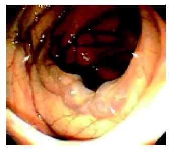
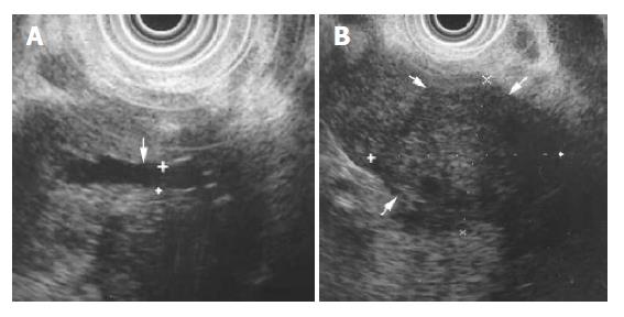
Physical examination revealed a chronically ill look, normal vital signs, no pale conjunctiva, no icteric sclera, soft and flat abdomen, neither hepatosplenomegaly nor palpable mass, and epigastric tenderness. The patient had received abdominal ultrasonography and esophagogas-troduodenoscopy (EGD) examinations that revealed nothing abnormal at a local medical clinic. Under the impression of colon cancer, the woman received colonoscopy at our outpatient clinic. Cecal diverticulum and isolated colon varices over the hepatic flexure were found (Figure 1). Under the suspicion of decompensated liver cirrhosis or pancreatic lesion, abdominal ultrasonography was repeated at our outpatient clinic. Abdominal ultrasonography revealed noncirrhotic liver and no evidence of portal hypertension, normal biliary trees, slightly dilated main pancreatic duct with 3 mm diameter over the pancreatic body, and poorly demonstrated pancreas head and tail area. EGD was also repeated to evaluate for the possibility of the combination of esophageal and gastric varices. Superficial gastritis was the only endoscopy finding. Upon directed endoscopic examination, no varices were seen in the esophagus, stomach and duodenum. Consequently, endoscopic ultrasonography (EUS) was conducted, and a dilated main pancreatic duct (2.7-3.6 mm in different sites) was detected, with a single hypoechoic mass (3.4×2.8 cm) over the uncinate pancreatic head (Figure 2). Abdominal computed tomography (CT), which was performed later, revealed a pancreatic head mass with mild pancreatic duct dilatation, no intra-abdominal metastasis, suspicious duodenal invasion, prominent collateral circulation over right side colon and occlusion of the superior mesenteric vein (Figure 3).
Because of suspected pancreatic head cancer, the patient was admitted for further study and treatment. Chest radiograph showed essentially negative findings in the lungs, mediastinum, heart, airway and diaphragm. Meanwhile, laboratory tests, including liver function and renal function tests, were normal but revealed raised amylase 298 IU/L (normal, 27-137 IU/L) and lipase 3137 IU/L (<170 IU/L) as well as mild anemia (hemoglobin 11.6 g/dL, 12-16 g/dL). Carcinoembryonic antigen (CEA) 1.6 ng/mL (<5 ng/mL) was normal but raised CA 19-9 with 53.1 IU/mL (<37 IU/mL) was noted. Duodenoscopy found no mucosal invasion by the pancreatic head cancer. Magnetic resonance cholangiopancreatography (MRCP) revealed mild dilatation of the pancreatic and common bile ducts, as well as a small filling defect over their junction. Magnetic resonance image of abdomen illustrated no definite intraabdominal metastatic lesion, although occlusion of the superior mesenteric vein was noted. EUS-guided fine needle aspiration (EUS-FNA) was conducted for cytological study, and revealed ductal epithelial hyperplasia and some cell necrosis without malignant cells.
Given the final impression of uncinate pancreatic head cancer without definite intra-abdominal metastasis, surgical laparotomy was arranged. During laparotomy, miliary metastasis was found over the liver surface, although the peritoneum was unaffected. Frozen sections of metastatic liver lesions displayed poorly differentiated adenocarcinoma. Pancreatic cancer with multiple liver metastasis was diagnosed, and since curative surgical resection was contraindicative, chemotherapy was selected as the treatment regimen.
Varices located over the esophagus and stomach are common complications of portal hypertension in the patients with advanced liver disease. Colon varices had been reported in patients diagnosed of liver cirrhosis, and most of them are located over the rectum only[3-5]. Varices over the right side colon among cirrhotic or noncirrhotic patients with portal hypertension were very rare[2]. There were several case reports of right side colon varices in the literature[2,6-16]. Causes of colon varices include congenital or familial venous anomalies, portal hypertension or occlusion, congestive heart failure, splenic vein thrombosis and mesenteric vein obstruction[2,9]. Cirrhotic or noncirrhotic portal hypertension is the most frequently encountered cause of colon varices[2-5]. Pancreatic cancer with mesenteric vein occlusion was never found as a cause of colon varices. In our report, isolated hepatic flexure colon varices were found incidentally. Under this finding of isolated colon varices over the hepatic flexure, most physicians easily reach a first impression of cirrhotic or noncirrhotic portal hypertension[2-5]. Other causes of colon varices may be missed or ignored if clinicians merely focus on common causes of colon varices, such as cirrhotic or noncirrhotic portal hypertension. In the case reported here, further study showed a novel cause of colon varices due to uncinate pancreatic head cancer with superior mesenteric vein occlusion.
Patients with pancreatic disease, including chronic pancreatitis or pancreatic cancer, may exhibit isolated gastric varices[17-21]. In 1987, Bradley reported 11 cases of splenic vein thrombosis caused by chronic pancreatitis[17]. One of the 11 patients displayed colon varices with bleeding, with the colon varices being located over the splenic flexure. While studying the present case, isolated hepatic flexure colon varices implied that the abdominal ultrasonography should be repeated owing to suspicions of advanced liver or pancreatic disease-induced venous thrombosis, the latter just like the pathogenesis of sinistral portal hypertension[17-21]. As is well known, it is difficult to show the pancreas via ultrasonography in certain situations. In this case, the patient received ultrasonography investigation at a local medical clinic. Repeated abdominal ultrasonography only revealed mild dilation of the pancreatic duct. This finding was trivial, but suggests that the pancreatic lesion contributed to the colon varices and symptoms. Subsequent investigations including EUS, abdominal CT scan and MRCP clearly demonstrated the pancreatic head mass with occlusion of the superior mesenteric vein. Most patients suffering from isolated gastric varices in chronic pancreatitis or pancreatic cancer have left-sided portal hypertension owing to occlusion of the splenic vein. In this patient with pancreatic head cancer, isolated hepatic flexure colon varices were caused by occlusion of the superior mesenteric vein. These two varieties of varices complicated by pancreatic disease share similar mechanisms but have different locations of venous occlusions. The cancer on the uncinate process of the pancreatic head possibly contributed to the development of superior mesenteric vein occlusion and did not cause splenic vein occlusion. The specific location of uncinate pancreatic cancer might cause delayed diagnosis, supplying sufficient time for the development of the colon varices. Miao et al[14], also reported one case of extensive colonic varices owing to mesenteric venous obstruction, but the cause in this case was ileal carcinoid tumor.
As early as in 1980, Izsak et al[2], had reviewed the literature and found three types of therapies of colon varices, including conservative, colonic resection and portocaval shunt. Transjugular intrahepatic portosystemic shunt (TIPS) that was an alternative method to reduce portal hypertension could get hemostasis or prevention of colon varices bleeding[13,22,23]. Since 1985, endoscopic sclerotherapy (EIS) had been performed successfully to stop rectosigmoid varices bleeding[24-27]. Endoscopic band ligation (EVL) had also been reported to control the rectal bleeding[28,29]. Combined EIS and EVL could control rectal varices bleeding successfully in one case report[30]. Another case report showed colon varices bleeding, which sigmoid resection had failed to control, was successfully managed by administration of somatostatin analog[31].
The case presented here illustrates a novel cause of colon varices. The special location of pancreatic cancer in uncinate process caused superior mesenteric vein occlusion resulting in the formation of isolated colon varices located over the hepatic flexure. In facing the finding of isolated hepatic flexure colon varices, pancreatic head cancer over the uncinate process with superior mesenteric vein invasion should be included in the table of differentiated diagnoses.
Science Editor Guo SY Language Editor Elsevier HK
| 1. | Feldman M, Smith VM, Warner CG. Varices of the colon. Report of three cases. JAMA. 1962;179:729-730. [RCA] [PubMed] [DOI] [Full Text] [Cited by in Crossref: 66] [Cited by in RCA: 59] [Article Influence: 0.9] [Reference Citation Analysis (0)] |
| 2. | Izsak EM, Finlay JM. Colonic varices. Three case reports and review of the literature. Am J Gastroenterol. 1980;73:131-136. [PubMed] |
| 3. | Rabinovitz M, Schade RR, Dindzans VJ, Belle SH, Van Thiel DH, Gavaler JS. Colonic disease in cirrhosis. An endoscopic evaluation in 412 patients. Gastroenterology. 1990;99:195-199. [PubMed] |
| 4. | Ganguly S, Sarin SK, Bhatia V, Lahoti D. The prevalence and spectrum of colonic lesions in patients with cirrhotic and noncirrhotic portal hypertension. Hepatology. 1995;21:1226-1231. [PubMed] |
| 5. | Bresci G, Gambardella L, Parisi G, Federici G, Bertini M, Rindi G, Metrangolo S, Tumino E, Bertoni M, Cagno MC. Colonic disease in cirrhotic patients with portal hypertension: an endoscopic and clinical evaluation. J Clin Gastroenterol. 1998;26:222-227. [RCA] [PubMed] [DOI] [Full Text] [Cited by in Crossref: 36] [Cited by in RCA: 33] [Article Influence: 1.2] [Reference Citation Analysis (0)] |
| 6. | Kori M, Keter D, Grunshpan M, Zimmerman J, Ackerman Z. Familial colonic varices. J Pediatr Gastroenterol Nutr. 2000;30:447-449. [RCA] [PubMed] [DOI] [Full Text] [Cited by in Crossref: 7] [Cited by in RCA: 9] [Article Influence: 0.4] [Reference Citation Analysis (0)] |
| 7. | Solis-Herruzo JA. Familial varices of the colon diagnosed by colonscopy. Gastrointest Endosc. 1977;24:85-86. [RCA] [PubMed] [DOI] [Full Text] [Cited by in Crossref: 23] [Cited by in RCA: 22] [Article Influence: 0.5] [Reference Citation Analysis (0)] |
| 8. | Morini S, Caruso F, De Angelis P. Familial varices of the small and large bowel. Endoscopy. 1993;25:188-190. [RCA] [PubMed] [DOI] [Full Text] [Cited by in Crossref: 10] [Cited by in RCA: 11] [Article Influence: 0.3] [Reference Citation Analysis (0)] |
| 9. | Atin V, Sabas JA, Cotano JR, Madariaga M, Galan D. Familial varices of the colon and small bowel. Int J Colorectal Dis. 1993;8:4-8. [RCA] [PubMed] [DOI] [Full Text] [Cited by in Crossref: 17] [Cited by in RCA: 18] [Article Influence: 0.6] [Reference Citation Analysis (0)] |
| 10. | Smith TR. CT demonstration of ascending colon varices. Clin Imaging. 1994;18:4-6. [RCA] [PubMed] [DOI] [Full Text] [Cited by in Crossref: 5] [Cited by in RCA: 6] [Article Influence: 0.2] [Reference Citation Analysis (0)] |
| 11. | Villarreal HA, Marts BC, Longo WE, Ure T, Vernava AM, Joshi S. Congenital colonic varices in the adult. Report of a case. Dis Colon Rectum. 1995;38:990-992. [RCA] [PubMed] [DOI] [Full Text] [Cited by in Crossref: 10] [Cited by in RCA: 11] [Article Influence: 0.4] [Reference Citation Analysis (0)] |
| 12. | Shrestha R, Dunkelberg JC, Schaefer JW. Idiopathic colonic varices: an unusual cause of massive lower gastrointestinal hemorrhage. Am J Gastroenterol. 1995;90:496-497. [PubMed] |
| 13. | Allgaier HP, Ochs A, Haag K, Hauenstein KH, Tittor W, Rössle M, Blum HE. Recurrent bleeding from colonic varices in portal hypertension. The successful prevention of recurrence by the implantation of a transjugular intrahepatic stent-shunt (TIPS). Dtsch Med Wochenschr. 1995;120:1773-1776. [RCA] [PubMed] [DOI] [Full Text] [Cited by in Crossref: 9] [Cited by in RCA: 11] [Article Influence: 0.4] [Reference Citation Analysis (0)] |
| 14. | Miao YM, Catnach SM, Barrison IG, O'Reilly A, Divers AR. Colonic variceal bleeding in a patient with mesenteric venous obstruction due to an ileal carcinoid tumour. Eur J Gastroenterol Hepatol. 1996;8:1133-1135. [RCA] [PubMed] [DOI] [Full Text] [Cited by in Crossref: 6] [Cited by in RCA: 7] [Article Influence: 0.2] [Reference Citation Analysis (0)] |
| 15. | Shaper KR, Jarmulowicz M, Dick R, Cuthbert ND, Davidson BR. Massive colonic haemorrhage from a solitary caecal varix. HPB Surg. 1996;9:253-256. [RCA] [PubMed] [DOI] [Full Text] [Full Text (PDF)] [Cited by in Crossref: 3] [Cited by in RCA: 4] [Article Influence: 0.1] [Reference Citation Analysis (0)] |
| 16. | Schmidt C, Klomp HJ, Doniec M, Grimm H. Primary varices of the colon. A rare cause of gastrointestinal bleeding. Dtsch Med Wochenschr. 1998;123:1069-1072. [RCA] [PubMed] [DOI] [Full Text] [Cited by in Crossref: 3] [Cited by in RCA: 4] [Article Influence: 0.1] [Reference Citation Analysis (0)] |
| 17. | Bradley EL. The natural history of splenic vein thrombosis due to chronic pancreatitis: indications for surgery. Int J Pancreatol. 1987;2:87-92. [PubMed] |
| 18. | Evans GR, Yellin AE, Weaver FA, Stain SC. Sinistral (left-sided) portal hypertension. Am Surg. 1990;56:758-763. [PubMed] |
| 19. | Sakorafas GH, Sarr MG, Farley DR, Farnell MB. The significance of sinistral portal hypertension complicating chronic pancreatitis. Am J Surg. 2000;179:129-133. [RCA] [PubMed] [DOI] [Full Text] [Cited by in Crossref: 111] [Cited by in RCA: 119] [Article Influence: 4.8] [Reference Citation Analysis (0)] |
| 20. | Chang CY. Pancreatic adenocarcinoma presenting as sinistral portal hypertension: an unusual presentation of pancreatic cancer. Yale J Biol Med. 1999;72:295-300. [PubMed] |
| 21. | Jaroszewski DE, Schlinkert RT, Gray RJ. Laparoscopic splenectomy for the treatment of gastric varices secondary to sinistral portal hypertension. Surg Endosc. 2000;14:87. [PubMed] |
| 22. | Shibata D, Brophy DP, Gordon FD, Anastopoulos HT, Sentovich SM, Bleday R. Transjugular intrahepatic portosystemic shunt for treatment of bleeding ectopic varices with portal hypertension. Dis Colon Rectum. 1999;42:1581-1585. [RCA] [PubMed] [DOI] [Full Text] [Cited by in Crossref: 95] [Cited by in RCA: 85] [Article Influence: 3.3] [Reference Citation Analysis (0)] |
| 23. | Chevallier P, Motamedi JP, Demuth N, Caroli-Bosc FX, Oddo F, Padovani B. Ascending colonic variceal bleeding: utility of phase-contrast MR portography in diagnosis and follow-up after treatment with TIPS and variceal embolization. Eur Radiol. 2000;10:1280-1283. [RCA] [PubMed] [DOI] [Full Text] [Cited by in Crossref: 14] [Cited by in RCA: 14] [Article Influence: 0.6] [Reference Citation Analysis (0)] |
| 24. | Wang M, Desigan G, Dunn D. Endoscopic sclerotherapy for bleeding rectal varices: a case report. Am J Gastroenterol. 1985;80:779-780. [PubMed] |
| 25. | Yamanaka T, Shiraki K, Ito T, Sugimoto K, Sakai T, Ohmori S, Takase K, Nakano T, Oohashi Y, Okuda Y. Endoscopic sclerotherapy (ethanolamine oleate injection) for acute rectal varices bleeding in a patient with liver cirrhosis. Hepatogastroenterology. 2002;49:941-943. [PubMed] |
| 26. | Iwase H, Kyogane K, Suga S, Morise K. Endoscopic ultrasonography with color Doppler function in the diagnosis of rectal variceal bleeding. J Clin Gastroenterol. 1994;19:227-230. [RCA] [PubMed] [DOI] [Full Text] [Cited by in Crossref: 13] [Cited by in RCA: 11] [Article Influence: 0.4] [Reference Citation Analysis (0)] |
| 27. | Chen WC, Hou MC, Lin HC, Chang FY, Lee SD. An endoscopic injection with N-butyl-2-cyanoacrylate used for colonic variceal bleeding: a case report and review of the literature. Am J Gastroenterol. 2000;95:540-542. [RCA] [PubMed] [DOI] [Full Text] [Cited by in Crossref: 37] [Cited by in RCA: 43] [Article Influence: 1.7] [Reference Citation Analysis (0)] |
| 28. | Uno Y, Munakata A, Ishiguro A, Fukuda S, Sugai M, Munakata H. Endoscopic ligation for bleeding rectal varices in a child with primary extrahepatic portal hypertension. Endoscopy. 1998;30:S107-S108. [RCA] [PubMed] [DOI] [Full Text] [Cited by in Crossref: 11] [Cited by in RCA: 9] [Article Influence: 0.3] [Reference Citation Analysis (0)] |
| 29. | Firoozi B, Gamagaris Z, Weinshel EH, Bini EJ. Endoscopic band ligation of bleeding rectal varices. Dig Dis Sci. 2002;47:1502-1505. [RCA] [PubMed] [DOI] [Full Text] [Cited by in Crossref: 31] [Cited by in RCA: 27] [Article Influence: 1.2] [Reference Citation Analysis (0)] |
| 30. | Shudo R, Yazaki Y, Sakurai S, Uenishi H, Yamada H, Sugawara K, Kohgo Y. Combined endoscopic variceal ligation and sclerotherapy for bleeding rectal varices associated with primary biliary cirrhosis: a case showing a long-lasting favorable response. Gastrointest Endosc. 2001;53:661-665. [RCA] [PubMed] [DOI] [Full Text] [Cited by in Crossref: 15] [Cited by in RCA: 15] [Article Influence: 0.6] [Reference Citation Analysis (0)] |
| 31. | Chakravarty BJ, Riley JW. Control of colonic variceal haemorrhage with a somatostatin analogue. J Gastroenterol Hepatol. 1996;11:305-306. [RCA] [PubMed] [DOI] [Full Text] [Cited by in Crossref: 10] [Cited by in RCA: 10] [Article Influence: 0.3] [Reference Citation Analysis (0)] |


