Published online Mar 28, 2005. doi: 10.3748/wjg.v11.i12.1859
Revised: May 12, 2004
Accepted: August 22, 2004
Published online: March 28, 2005
AIM: To tattoo gastric mucosa with a novel medical device which could be used to monitor and follow-up gastric mucosal lesions.
METHODS: Combining endoscopic biopsy with sclerotherapy injection, we designed a new device that could perform biopsy and injection simultaneously. We performed endoscopies on a pig by using a novel endoscope tattoo biopsy forceps for 15 mo. At the same time, we used two-step method combining sclerotherapy injection needle with endoscopic biopsy. The acuity, inflammation and duration of endoscopy were compared between two methods.
RESULTS: Compared with the old two-step method, although the inflammation induced by our new device was similar, the duration of procedure was markedly decreased and the acuity of tattooing was better than the old two-step method. All characteristics of the novel device complied with national safety guidelines. Follow-up gastroscopy after 15 mo showed the stained site with injection of 1:100 0.5 mL of India ink was still markedly visible with little inflammatory reaction.
CONCLUSION: Endoscopic tattooing biopsy forceps can be widely used in monitoring precancerous lesions. Its safety and effectiveness has been established in animals.
- Citation: Si JM, Sun LM, Fan YJ, Wang LJ. Trial of a novel endoscopic tattooing biopsy forceps on animal model. World J Gastroenterol 2005; 11(12): 1859-1861
- URL: https://www.wjgnet.com/1007-9327/full/v11/i12/1859.htm
- DOI: https://dx.doi.org/10.3748/wjg.v11.i12.1859
Fiber-optic endoscopy is widely used to diagnose gastrointestinal diseases, including chronic inflammation, precancerous lesion, benign and malignant tumors, polyps and congenital malformations. As an important medical diagnostic modality, endoscopic biopsy allows a pathologic diagnosis. This is especially useful in monitoring precancerous lesions, pre- or post-operation follow-up[1,2]. However, identifying and demarcating the suspicious area for follow-up remain problematic[3-6]. One method to solve this problem is to use an injection needle for mucosa tattooing, then routine endoscopic biopsy was performed for pathologic inspection. This technique is not only cumbersome, but also may lack precision. In this regard, tattooing biopsy forceps that permit simultaneous marking and biopsy of the suspicious lesion during the same procedure may be very useful.
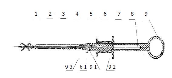
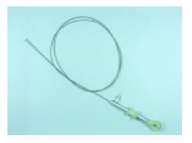
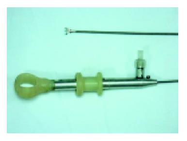
The structure of endoscopic tattooing biopsy forceps is shown in Figures 1, 2, and 3. It consists of 9 parts: (1) a syringe needle, on which a threaded hole was made for fixation guide wire of the biopsy forceps; (2) a bowl-shaped notched biopsy forceps, which can be fixed on mucosa for biopsy without damaging the needle; (3) a fixation sleeve of biopsy forceps, which is provided with a lumen to allow the guide wire to move freely; (4) a biopsy forceps guide wire; (5) a metallic tube; (6) a transfusion tube which is provided with syringe nozzle 6-1; (7) a control block of syringe needle and biopsy forceps on which an adaptor for piece 8 connecting the transfusion tube can be fixed; (8) an adaptor piece of transfusion tube, which drives the transfusion tube to shift leftwards or rightwards and in turn drives the needle to move forwards or backwards and the biopsy forceps to open or close; (9) a pull-rod shank, on which there is a slot of syringe nozzle 9-1, slot for pull-rod 9-2 (to make the transfusion tube shift leftwards or rightwards freely within a given range) and handle 9-3.
We performed gastroscopic tattooing biopsies of gastric mucosa on one pig, using medical India ink (Pelikan, Hanower, Germany) for staining. India ink was diluted and sterilized before use[7]. The pig was subjected to fiber-optic gastroscopy under general anesthesia and gastric mucosa was biopsied with both the novel tattooing biopsy forceps and the old methods on different sites of gastric mucosa. Then the safety and effectiveness of the tattooing biopsy were checked, several follow-up gastroscopies were performed at the intervals of 1, 3 d, 1 wk, 1, 3, 6, 12, 15 mo respectively. The concentration of India ink was 1:10, 1:100, 1:1000 respectively, and the dosage of injection was 0.3 mL, 0.5 mL, and 0.7 mL respectively. The acuity of tattooing was described as three scores: 0, nothing could be seen; 1, tattooing could be seen indistinctly; 2, tattooing was clearly seen. The inflammation induced by biopsy was described as four scores: 0, no inflammation, tattooing and impact on observation; 1, mild inflammation, tattooing and impact on observation; 2, moderate inflammation, tattooing and impact on observation; 3, severe inflammation, tattooing and impact on observation. The duration of each procedure was recorded and compared. Twelve biopsy specimens were obtained with each novel tattooing biopsy forceps and ordinary biopsy forceps. Tissue samples were examined for the following parameters: weight (mg), size (mm3), depth, and adequacy of the specimens for histologic information (0, inadequate; 1, suboptimal; and 2, adequate)[8].
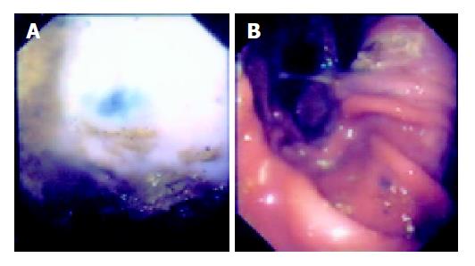
No complication was noted during 15-mo follow-up. Follow-up gastroscopy after 15 mo showed with injection of 0.5 mL (1:10) of India ink, the dye site was still visible with little inflammatory reaction (Table 1, Figure 4). A total of 24 tissue samples were available for analysis. As far as the inflammation due to tattooing was concerned, inflammation induced by the novel tattooing biopsy forceps method and the old two-step methods was similar. The acuity of tattooing was better in novel tattooing biopsy forceps method than in the old two-step method. The mean time of endoscopy with novel tattooing biopsy forceps were significantly short (40 s) compared to the traditional two-step method (70 s). Overall, both forceps provided specimens with a similar size and depth. When the adequacy of specimens was assessed for histologic diagnosis, no significant difference was noted between two kinds of forceps.
| Concentration and dosage | Acuity of tattooing | Inflammation of tattooing |
| 1:10, 0.3 mL | 1 | 1 |
| 1:10, 0.5 mL | 2 | 1 |
| 1:10, 0.7 mL | 2 | 2 |
| 1:100, 0.3 mL | 1 | 2 |
| 1:100, 0.5 mL | 1 | 1 |
| 1:100, 0.7 mL | 1 | 2 |
| 1:1000, 0.3 mL | 0 | 0 |
| 1:1000, 0.5 mL | 0 | 0 |
| 1:1000, 0.7 mL | 1 | 1 |
Tissue staining is used as an adjunctive technique during gastrointestinal endoscopy. Chemical agents have been applied to the detection of mucosal surface characteristics of the gastrointestinal epithelium[9-11]. This aids in the recognition of subtle lesions or allows direct targeting of biopsies to increase the endoscopic diagnostic accuracy. Traditional method was performed by using an injection needle for mucosa tattooing, then routine endoscopic biopsy was carried out for pathologic inspection. But this technique is not only cumbersome, but also may lack precision. It takes a long time and easy to miss the targeting site. The novel tattooing biopsy forceps that permits simultaneous marking and biopsy of the suspicious lesion during the same procedure is very useful. By using it, the distress of patients during endoscopy could be reduced. In addition, it may avoid the difficulty in identifying the area of previous biopsies with follow-up examinations. If no marking was made for minor pathological changes after biopsy it might be difficult to identify the suspicious area during surgery. This could preclude effective treatment and correct assessment of the effect of chemotherapy. Since this device is a disposable tattooing biopsy forceps, cross contamination and infection can be prevented. It can be used for inspection of precancerous lesions with a long-term follow-up of various mucosa lesions in the gastrointestinal tract, genitourinary tract and tracheobronchial tree.
India ink, a colloidal suspension of carbon particles, is most widely used as a tattooing agent. The carbon is derived from incomplete combustion of petroleum products in an oxygen-depleted system. Carbon tattooing has been used for decades to tattoo the skin without toxicity, and India ink tattoos of the gastrointestinal tract have be reported to be similarly safe[4,12-14]. A 0.5 mL of India ink solution diluted to 1:10 with sterile saline seems to be the best choice.
There are two conduits (one for the biopsy guide wire, the other for the transfusion tube) inside the cavity of tattooing biopsy forceps. Hence it has strict requirements on the technological process. Through a series of experiments, we at last decided to use a stainless steel wire with a diameter of 0.28 mm and a transfusion tube with an outside diameter of 0.75 mm and an inside diameter of 0.25 mm, which prevented the obstruction of the transfusion tube and allowed a maximum flexibility of the closing/opening of the biopsy bowl. This tattooing biopsy forceps is a practical medical device and has been used in chinese patients (ZL 01 2 76535.X).
The endoscopic tattooing biopsy forceps can be widely used in monitoring precancerous lesions. Its safety and effectiveness has been established in animals. Further studies are needed to assess its therapeutic impact and safety.
We thank Dr. David Mc Fadden, Loma Linda University, USA, for reviewing our article and making corrections.
| 1. | Wan J, Zhang ZQ, Zhu C, Wang MW, Zhao DH, Fu YH, Zhang JP, Wang YH, Wu BY. Colonoscopic screening and follow-up for colorectal cancer in the elderly. World J Gastroenterol. 2002;8:267-269. [PubMed] |
| 2. | Noguchi Y, Yoshikawa T, Tsuburaya A, Motohashi H, Karpeh MS, Brennan MF. Is gastric carcinoma different between Japan and the United States? Cancer. 2000;89:2237-2246. [RCA] [PubMed] [DOI] [Full Text] [Cited by in RCA: 4] [Reference Citation Analysis (0)] |
| 3. | McArthur CS, Roayaie S, Waye JD. Safety of preoperation endoscopic tattoo with india ink for identification of colonic lesions. Surg Endosc. 1999;13:397-400. [RCA] [PubMed] [DOI] [Full Text] [Cited by in Crossref: 60] [Cited by in RCA: 61] [Article Influence: 2.3] [Reference Citation Analysis (0)] |
| 4. | Lane KL, Vallera R, Washington K, Gottfried MR. Endoscopic tattoo agents in the colon. Tissue responses and clinical implications. Am J Surg Pathol. 1996;20:1266-1270. [RCA] [PubMed] [DOI] [Full Text] [Cited by in Crossref: 51] [Cited by in RCA: 52] [Article Influence: 1.8] [Reference Citation Analysis (0)] |
| 5. | Price N, Gottfried MR, Clary E, Lawson DC, Baillie J, Mergener K, Westcott C, Eubanks S, Pappas TN. Safety and efficacy of India ink and indocyanine green as colonic tattooing agents. Gastrointest Endosc. 2000;51:438-442. [RCA] [PubMed] [DOI] [Full Text] [Cited by in Crossref: 70] [Cited by in RCA: 79] [Article Influence: 3.2] [Reference Citation Analysis (0)] |
| 6. | Shaffer RT, Francis JM, Carrougher JG, Root SS, Angueira CE, Szyjkowski R, Kadakia SC. India ink tattooing in the esophagus. Gastrointest Endosc. 1998;47:257-260. [RCA] [PubMed] [DOI] [Full Text] [Cited by in Crossref: 20] [Cited by in RCA: 21] [Article Influence: 0.8] [Reference Citation Analysis (0)] |
| 7. | Salomon P, Berner JS, Waye JD. Endoscopic India ink injection: a method for preparation, sterilization, and administration. Gastrointest Endosc. 1993;39:803-805. [RCA] [PubMed] [DOI] [Full Text] [Cited by in Crossref: 36] [Cited by in RCA: 33] [Article Influence: 1.0] [Reference Citation Analysis (0)] |
| 8. | Woods KL, Anand BS, Cole RA, Osato MS, Genta RM, Malaty H, Gurer IE, Rossi DD. Influence of endoscopic biopsy forceps characteristics on tissue specimens: results of a prospective randomized study. Gastrointest Endosc. 1999;49:177-183. [RCA] [PubMed] [DOI] [Full Text] [Cited by in Crossref: 39] [Cited by in RCA: 39] [Article Influence: 1.5] [Reference Citation Analysis (0)] |
| 9. | Dawsey SM, Fleischer DE, Wang GQ, Zhou B, Kidwell JA, Lu N, Lewin KJ, Roth MJ, Tio TL, Taylor PR. Mucosal iodine staining improves endoscopic visualization of squamous dysplasia and squamous cell carcinoma of the esophagus in Linxian, China. Cancer. 1998;83:220-231. [RCA] [PubMed] [DOI] [Full Text] [Cited by in RCA: 6] [Reference Citation Analysis (0)] |
| 10. | Hiki Y. Endoscopic diagnosis of mucosal cancer. Semin Surg Oncol. 1999;17:91-95. [RCA] [PubMed] [DOI] [Full Text] [Cited by in RCA: 1] [Reference Citation Analysis (0)] |
| 11. | Namihisa A, Miwa H, Watanabe H, Kobayashi O, Ogihara T, Sato N. A new technique: light-induced fluorescence endoscopy in combination with pharmacoendoscopy. Gastrointest Endosc. 2001;53:343-348. [RCA] [PubMed] [DOI] [Full Text] [Cited by in Crossref: 11] [Cited by in RCA: 12] [Article Influence: 0.5] [Reference Citation Analysis (0)] |
| 12. | Hyman N, Waye JD. Endoscopic four quadrant tattoo for the identification of colonic lesions at surgery. Gastrointest Endosc. 1991;37:56-58. [RCA] [PubMed] [DOI] [Full Text] [Cited by in Crossref: 54] [Cited by in RCA: 49] [Article Influence: 1.4] [Reference Citation Analysis (0)] |
| 13. | Nizam R, Siddiqi N, Landas SK, Kaplan DS, Holtzapple PG. Colonic tattooing with India ink: benefits, risks, and alternatives. Am J Gastroenterol. 1996;91:1804-1808. [PubMed] |
| 14. | Shatz BA, Weinstock LB, Swanson PE, Thyssen EP. Long-term safety of India ink tattoos in the colon. Gastrointest Endosc. 1997;45:153-156. [RCA] [PubMed] [DOI] [Full Text] [Cited by in Crossref: 50] [Cited by in RCA: 54] [Article Influence: 1.9] [Reference Citation Analysis (0)] |


