Published online Mar 14, 2005. doi: 10.3748/wjg.v11.i10.1532
Revised: August 15, 2004
Accepted: October 18, 2004
Published online: March 14, 2005
AIM: To determine the efficacy of multislice CT for gastroenteric and hepatic surgery.
METHODS: Dual-phase helical computed tomography was performed in 50 of 51 patients who underwent gastroenteric and hepatic surgeries. Twenty-eight, eighteen and four patients suffering from colorectal cancer, gastric cancer, and liver cancer respectively underwent colorectal surgery (laparoscopic surgery: 6 cases), gastrectomy, and hepatectomy. Three-dimensional computed tomography imaging of the inferior mesenteric artery, celiac artery and hepatic artery was performed. And in the follow-up examination of postoperative patients, multiplanar reconstruction image was made in case of need.
RESULTS: Scans in 50 patients were technically satisfactory and included in the analysis. Depiction of major visceral arteries, which were important for surgery and other treatments, could be done in all patients. Preoperative visualization of the left colic artery and sigmoidal arteries, the celiac artery and its branches, and hepatic artery was very useful to lymph node dissection, the planning of a reservoir and hepatectomy. And multiplanar reconstruction image was helpful to diagnosis for the postoperative follow-up of patients.
CONCLUSION: Three-dimensional volume rendering or multiplanar reconstruction imaging performed by multislice computed tomography was very useful for gastroenteric and hepatic surgeries.
- Citation: Ohtani H, Kawajiri H, Arimoto Y, Ohno K, Fujimoto Y, Oba H, Adachi K, Hirano M, Terakawa S, Tsubakimoto M. Efficacy of multislice computed tomography for gastroenteric and hepatic surgeries. World J Gastroenterol 2005; 11(10): 1532-1534
- URL: https://www.wjgnet.com/1007-9327/full/v11/i10/1532.htm
- DOI: https://dx.doi.org/10.3748/wjg.v11.i10.1532
Computed tomography (CT) is one of the most useful imaging techniques for the evaluation of original and metastatic lesions in gastroenteric and hepatic cancer. Contrast-enhanced CT (CECT) in particular has extraordinary precision. At our hospital, CECT is routinely performed to screen intra-abdominal malignancies. Recently, a helical CT scanner was developed, which permits scanning of the whole abdomen in a single breath-hold in each scanning phase. Multislice helical CT, which performs multiplanar reconstruction (MPR), maximum intensity projection (MIP), and shaded surface display (SSD), was first used at our institution in November of 2002[1,2]. Arterial phase helical CT can immediately display the major visceral arteries, such as the celiac artery (CA), superior mesenteric artery (SMA), inferior mesenteric artery (IMA), and hepatic artery (HA)[3-5]. Preoperative visualization of these vessels is useful for lymph node dissection, preservation of the left colic artery (LCA), planning of the reservoir, and hepatectomy.
Dual-phase helical CT was performed with a high-speed scanner (Aquilion M8, Toshiba Medical Systems Co., Ltd., Tokyo, Japan) on 50 patients (28 male and 22 female) between December of 2002 and July of 2004. The ages of the patients ranged from 36 to 81 years (mean 65.9). One patient was unable to undergo CECT due to renal dysfunction. Twenty-eight, eighteen and four patients suffering from colorectal cancer, gastric cancer and liver cancer respectively underwent colorectal surgery (laparoscopic surgery: 6 cases), gastrectomy and hepatectomy respectively. A 20-gauge cannula was inserted into an antecubital and dual-phase helical CT was then performed after power injection of 1.5 mL/kg of iohexol at a rate of 3 mL/s. An automatic bolus-tracking program (Surestart, Toshiba Medical) was used to automatically start the arterial phase scan after the injection of iohexol. The CT values of the region of interest specified in the abdominal aorta were automatically calculated. The trigger for starting the diagnostic scan was set at an increase in aortic enhancement of 50 H, and the arterial phase helical CT scan started automatically at 17 s after the trigger level had been reached. Late phase scanning was initiated at 60 s after the arterial phase scan[6]. After the state of the original region was estimated, metastatic or other lesions were examined. During each phase, scanning was performed in a single breath-hold. Dual-phase helical CT data were then transferred to a Zio M900 workstation (ZioSoft, Inc., Morgan Hill, CA, USA), and three-dimensional CT (3DCT) images of IMA, CA or HA were reconstructed. Bones, large untargeted vessels and background tissues could be eliminated from the image. If necessary, the MPR or MIP image was reconstructed.
Dual phase helical CT is often performed to follow up postoperative patients, too. It is useful in diagnosis and screening.
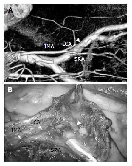
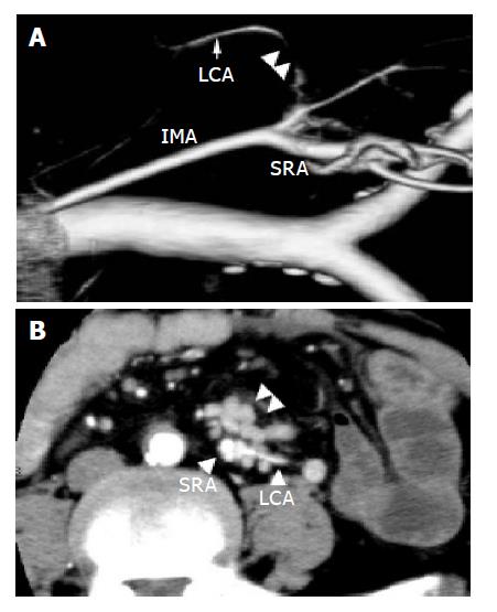
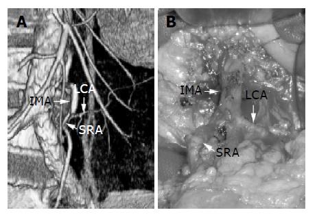
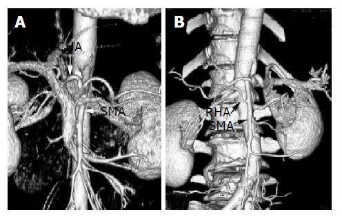
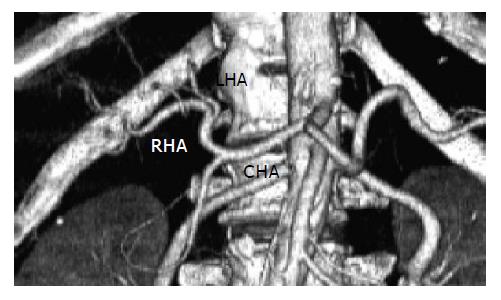
Before surgery for sigmoid colon or rectal cancer, 3DCT of the IMA was performed for dissection of the lymph nodes around the root of the IMA. In all patients except two, the LCA was easily preserved in open and laparoscopic surgery (Figure 1). In the two cases, the root of the IMA was resected because macroscopically metastatic lymph nodes adhered to the LCA. The adhesion had been preoperatively identified on 3DCT (Figure 2) and was confirmed in intraoperative findings. In one patient, a slight deformity of the SRA was noticed (Figure 3), but preoperative 3DCT enabled us to recognize the superior rectal artery (SRA) and LCA easily. In gastrectomy for gastric cancer, CT imaging was useful in examining the variation of the major vessels. Preoperative visualization of the common hepatic artery (CHA) diverging from the SMA (Figure 4A), and of the right hepatic artery (RHA) branching from the SMA (Figure 4B) was extremely helpful in lymph node dissection and in the planning of a reservoir in cases of multiple metastatic liver cancer. In hepatectomy, 3DCT was of particular use in a patient who could not undergo angiography due to severe arteriosclerosis (Figure 5).
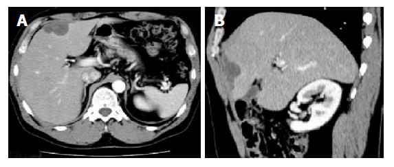
It was difficult to distinguish in the axial view whether a patient who underwent right hemicolectomy for ascending colon cancer suffered from dissemination or liver metastases (Figure 6A), however, he was diagnosed with dissemination by MPR image (Figure 6B).
Multislice CT has many merits including the short scanning time[7,8], ease of data acquisition for 3D volume rendering, MPR, SSD and MIP performance. In gastroenteric and hepatic surgery, multislice CT is highly useful, particularly for preoperative visualization of the regional arteries, and in gastroenteric surgery visualization of the major visceral arteries is extremely helpful in lymph node dissection in open and laparoscopic surgery. Until recently, preoperative definition of the vascular anatomy was achieved by conventional catheter angiography. However, at our hospital, dual-phase helical CT during the arterial and late phases of contrast material enhancement is now performed before gastroenteric and hepatic surgery. In resection of advanced sigmoid colon and rectal cancer, we routinely dissect the lymph nodes around the root of the IMA while preserving the LCA because resection of the root of the IMA occasionally causes ischemia of the oral side of the sigmoid colon, sometimes leading to anastomotic leakage[9]. In gastrectomy, preoperative imaging of arterial anomalies such as the anomalous RHA or the CHA arising from the SMA is helpful in lymph node dissection and in planning the reservoir in cases of metastatic liver cancer. It is important to recognize the anomalous left hepatic artery arising from the left gastric artery in lymph node dissection, and in hepatic surgery, imaging of the HA is important. 3DCT is especially useful in patients who cannot undergo angiography due to certain conditions such as severe arteriosclerosis.
Intra-abdominal lesions are screened in the axial view by dual-phase helical CT at our hospital. In some cases, additional MPR such as sagittal or coronal view is helpful to identify the specific condition of a tumor.
In the present study, we found no disagreement between 3DCT images and findings upon operation. 3DCT is able to show exactly the surrounding vascular structures and it is minimally invasive compared to conventional catheter angiography[10]. 3D volume-rendered image reconstruction such as MPR, SSD and MIP is easily and immediately acquired after the axial CT data are transferred to the Zio M900 workstation. 3DCT is a relatively simple technique, which provides many advantages, and dual-phase helical CT is useful not only in diagnosis and screening, but also in follow-up examination of postoperative patients.
In the future, 3DCT will prove to be essential in gastroenteric and hepatic surgeries.
Edited by Guo SY Language Editor Elsevier HK
| 1. | Hu H. Multi-slice helical CT: scan and reconstruction. Med Phys. 1999;26:5-18. [RCA] [PubMed] [DOI] [Full Text] [Cited by in Crossref: 369] [Cited by in RCA: 274] [Article Influence: 10.5] [Reference Citation Analysis (0)] |
| 2. | Hong KC, Freeny PC. Pancreaticoduodenal arcades and dorsal pancreatic artery: comparison of CT angiography with three-dimensional volume rendering, maximum intensity projection, and shaded-surface display. AJR Am J Roentgenol. 1999;172:925-931. [RCA] [PubMed] [DOI] [Full Text] [Cited by in Crossref: 48] [Cited by in RCA: 41] [Article Influence: 1.6] [Reference Citation Analysis (0)] |
| 3. | Chong M, Freeny PC, Schmiedl UP. Pancreatic arterial anatomy: depiction with dual-phase helical CT. Radiology. 1998;208:537-542. [RCA] [PubMed] [DOI] [Full Text] [Cited by in Crossref: 42] [Cited by in RCA: 46] [Article Influence: 1.7] [Reference Citation Analysis (0)] |
| 4. | Winter TC, Freeny PC, Nghiem HV, Hommeyer SC, Barr D, Croghan AM, Coldwell DM, Althaus SJ, Mack LA. Hepatic arterial anatomy in transplantation candidates: evaluation with three-dimensional CT arteriography. Radiology. 1995;195:363-370. [RCA] [PubMed] [DOI] [Full Text] [Cited by in Crossref: 108] [Cited by in RCA: 106] [Article Influence: 3.5] [Reference Citation Analysis (0)] |
| 5. | Smith PA, Klein AS, Heath DG, Chavin K, Fishman EK. Dual-phase spiral CT angiography with volumetric 3D rendering for preoperative liver transplant evaluation: preliminary observations. J Comput Assist Tomogr. 1998;22:868-874. [RCA] [PubMed] [DOI] [Full Text] [Cited by in Crossref: 35] [Cited by in RCA: 35] [Article Influence: 1.3] [Reference Citation Analysis (0)] |
| 6. | Kim T, Murakami T, Hori M, Takamura M, Takahashi S, Okada A, Kawata S, Cruz M, Federle MP, Nakamura H. Small hypervascular hepatocellular carcinoma revealed by double arterial phase CT performed with single breath-hold scanning and automatic bolus tracking. AJR Am J Roentgenol. 2002;178:899-904. [RCA] [PubMed] [DOI] [Full Text] [Cited by in Crossref: 56] [Cited by in RCA: 59] [Article Influence: 2.6] [Reference Citation Analysis (0)] |
| 7. | Heiken JP, Brink JA, Vannier MW. Spiral (helical) CT. Radiology. 1993;189:647-656. [RCA] [PubMed] [DOI] [Full Text] [Cited by in Crossref: 222] [Cited by in RCA: 172] [Article Influence: 5.4] [Reference Citation Analysis (0)] |
| 8. | Zeman RK, Fox SH, Silverman PM, Davros WJ, Carter LM, Griego D, Weltman DI, Ascher SM, Cooper CJ. Helical (spiral) CT of the abdomen. AJR Am J Roentgenol. 1993;160:719-725. [RCA] [PubMed] [DOI] [Full Text] [Cited by in Crossref: 110] [Cited by in RCA: 89] [Article Influence: 2.8] [Reference Citation Analysis (0)] |
| 9. | Okuda J, Tanigawa N. Laparoscopic surgery for rectal and sigmoid colon cancer. Nihon Rinsho. 2003;61 Suppl 7:391-395. [PubMed] |
| 10. | Bluemke DA, Chambers TP. Spiral CT angiography: an alternative to conventional angiography. Radiology. 1995;195:317-319. [RCA] [PubMed] [DOI] [Full Text] [Cited by in Crossref: 81] [Cited by in RCA: 61] [Article Influence: 2.0] [Reference Citation Analysis (0)] |


