Copyright
©The Author(s) 2003.
World J Gastroenterol. Jun 15, 2003; 9(6): 1361-1364
Published online Jun 15, 2003. doi: 10.3748/wjg.v9.i6.1361
Published online Jun 15, 2003. doi: 10.3748/wjg.v9.i6.1361
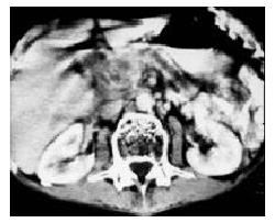
Figure 1 Mass with heterogeneous hypodensity located in the head of pancreas.
The body and tail of the pancreas appeared atrophy, and with enlarged main pancreatic duct.
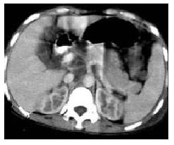
Figure 2 Pancreas appeared swollen diffusely with heterogeneous enhancement and vague margin.
A circle lymph nodes showed enhancement under pancreas.
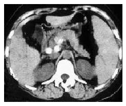
Figure 3 Calcification of pancreatic TB occurred under the body of the pancreas and retroperitoneal lymph nodes.
- Citation: Xia F, Poon RTP, Wang SG, Bie P, Huang XQ, Dong JH. Tuberculosis of pancreas and peripancreatic lymph nodes in immunocompetent patients: experience from China. World J Gastroenterol 2003; 9(6): 1361-1364
- URL: https://www.wjgnet.com/1007-9327/full/v9/i6/1361.htm
- DOI: https://dx.doi.org/10.3748/wjg.v9.i6.1361


