Copyright
©The Author(s) 2003.
World J Gastroenterol. Jun 15, 2003; 9(6): 1278-1281
Published online Jun 15, 2003. doi: 10.3748/wjg.v9.i6.1278
Published online Jun 15, 2003. doi: 10.3748/wjg.v9.i6.1278
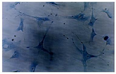
Figure 1 The hepatic stellate cells stained with toluidine after cultured for 10 d after isolation (× 66).
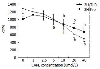
Figure 2 Effects of CAPE on 3H-TdR and 3H-proline uptake by HSCs.
aP < 0.05; bP < 0.01 vs groups without CAPE treatment.
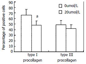
Figure 3 Effect of CAPE on type I and III procollagen gene expression.
aP < 0.05 vs the group without CAPE treatment.
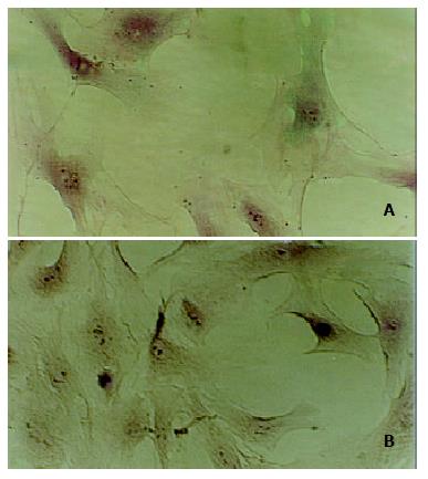
Figure 4 Apoptotic HSCs detected by TUNEL without (A) or with 40 μmol/L CAPE treatment (B).
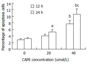
Figure 5 Effects of CAPE on HSC apoptosis.
aP < 0.05; bP < 0.01 vs the groups without CAPE treatment; cP < 0.01 vs the group with 40 μmol/L CAPE treated for 12 h.
-
Citation: Zhao WX, Zhao J, Liang CL, Zhao B, Pang RQ, Pan XH. Effect of caffeic acid phenethyl ester on proliferation and apoptosis of hepatic stellate cells
in vitro . World J Gastroenterol 2003; 9(6): 1278-1281 - URL: https://www.wjgnet.com/1007-9327/full/v9/i6/1278.htm
- DOI: https://dx.doi.org/10.3748/wjg.v9.i6.1278


