Copyright
©The Author(s) 2003.
World J Gastroenterol. Apr 15, 2003; 9(4): 670-673
Published online Apr 15, 2003. doi: 10.3748/wjg.v9.i4.670
Published online Apr 15, 2003. doi: 10.3748/wjg.v9.i4.670
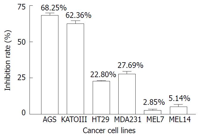
Figure 1 Effect of AR on the growth of 6 cancer cell lines.
Cells were treated with AR at the concentration of 100 µg/mL for 48 hrs and cell growth was measured with MTS method.
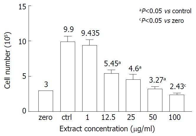
Figure 2 Effect of AR on AGS viability.
AGS cells were treated with different concentrations of AR for 48 hrs and cell viability was measured with trypan blue. At or below the concentra-tion of 50 µg/mL, AR was not cytotoxic to AGS cells. But at the concentration of 100 µg/mL, it showed cytotoxicity on AGS.
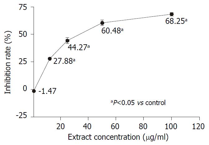
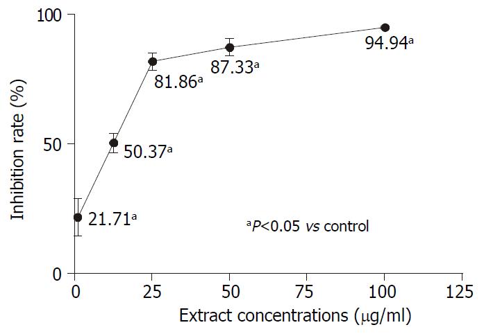
Figure 3 Dose-dependent effect of AR on AGS growth.
AGS cells were treated with a serial of diluted concentrations (1, 12.5, 25, 50 and 100 µg/mL) of AR for 48 hrs and cell growth was measured with MTS method. The inhibition effect of AR was improved accompanied by increase of concentration.
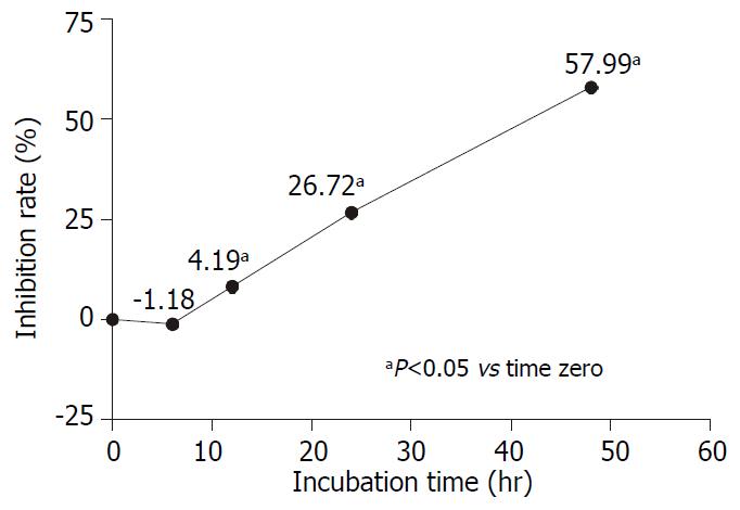
Figure 4 Time-dependent effect of AR on AGS growth.
AGS cells were treated with 50 µg/mL of AR for 6, 12, 24 and 48 hrs. The cell growth was measured by MTS method. The inhibition effect of AR on AGS growth was enhanced with incubation time.
Figure 5 Effect of AR on DNA synthesis of AGS.
AGS cells were treated with a serial of diluted concentrations (1, 12.5, 25, 50 and 100 µg/mL) of AR for 48 hrs and cell growth was mea-sured with tritium labeled thymidine incorporation assay. AR started to inhibit the DNA synthesis of AGS at 1 µg/mL and the inhibiting effect was dose-dependent.
- Citation: Lin J, Dong HF, Oppenheim J, Howard O. Effects of astragali radix on the growth of different cancer cell lines. World J Gastroenterol 2003; 9(4): 670-673
- URL: https://www.wjgnet.com/1007-9327/full/v9/i4/670.htm
- DOI: https://dx.doi.org/10.3748/wjg.v9.i4.670


