Copyright
©The Author(s) 2003.
World J Gastroenterol. Mar 15, 2003; 9(3): 627-630
Published online Mar 15, 2003. doi: 10.3748/wjg.v9.i3.627
Published online Mar 15, 2003. doi: 10.3748/wjg.v9.i3.627
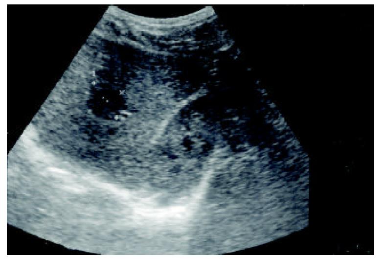
Figure 1 A hypoechoic lesion in the right lobe of the liver, segment S6, 24 × 22 mm in diameter (asterisks).
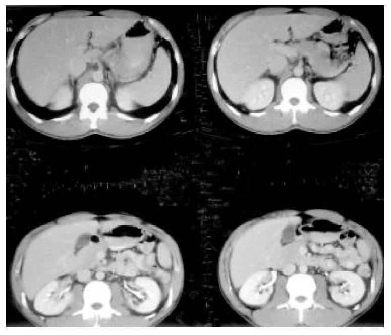
Figure 2 No evidence of hepatocellular tumor seen on upper abdominal CT with contrast.
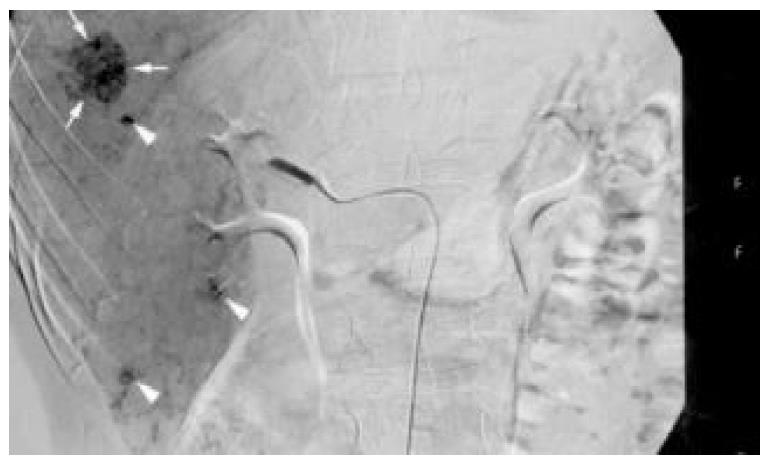
Figure 3 On angiography, there was an oval hypervascular lesion (black arrows) and multiple scattered tiny hypervascular spots in the right lobe of the liver (white arrows).
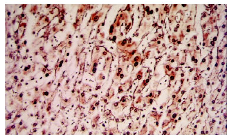
Figure 4 Slightly disarrayed hepatocytes with nuclear dyspla-sia were found.
Besides one-to-two layer reticulin stain, not consistent with HCC, was present. (Reticulin stain, × 250).
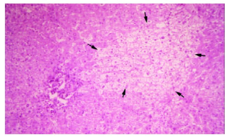
Figure 5 An oval-shape nodule (black arrows) which consisted of disarrayed hepatocytes with nuclear change and clear change in the cytoplasm was surrounded by normal liver tissue.
(H&E stain, × 125).
- Citation: Hsu CY, Chu CH, Lin SC, Yang FS, Yang TL, Chang KM. Concomitant hepatocellular adenoma and adenomatous hyperplasia in a patient without cirrhosis. World J Gastroenterol 2003; 9(3): 627-630
- URL: https://www.wjgnet.com/1007-9327/full/v9/i3/627.htm
- DOI: https://dx.doi.org/10.3748/wjg.v9.i3.627


