Copyright
©The Author(s) 2003.
World J Gastroenterol. Mar 15, 2003; 9(3): 516-520
Published online Mar 15, 2003. doi: 10.3748/wjg.v9.i3.516
Published online Mar 15, 2003. doi: 10.3748/wjg.v9.i3.516
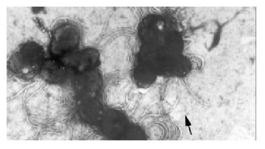
Figure 1 The flagella of coccoid H.
pylori under transmission electron microscope ×6000.
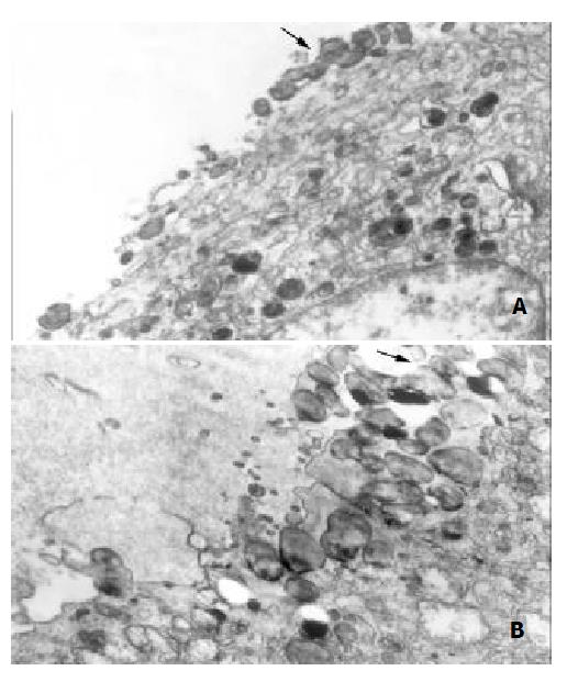
Figure 2 H.
pylori colonization in mouse stomach under trans-mission electron microscope. A. infection of spiral Hp. ×7 000; B. infection of coccoid Hp. ×9 000.
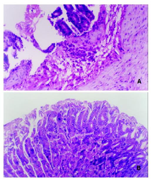
Figure 3 Light microscopy for gastric mucosa of mice.
H&E×200. A. infection of spiral Hp; B. infection of coccoid Hp.
-
Citation: She FF, Lin JY, Liu JY, Huang C, Su DH. Virulence of water-induced coccoid
Helicobacter pylori and its experimental infection in mice. World J Gastroenterol 2003; 9(3): 516-520 - URL: https://www.wjgnet.com/1007-9327/full/v9/i3/516.htm
- DOI: https://dx.doi.org/10.3748/wjg.v9.i3.516


