Copyright
©The Author(s) 2003.
World J Gastroenterol. Mar 15, 2003; 9(3): 399-403
Published online Mar 15, 2003. doi: 10.3748/wjg.v9.i3.399
Published online Mar 15, 2003. doi: 10.3748/wjg.v9.i3.399
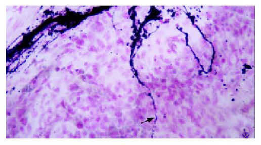
Figure 1 GAL-immunostaining of esophageal carcinoma.
Scat-tered nerve fibers (arrows) branched almost vertically from nerve bundles in connective tissue and entered into tumor parenchyma × 250.
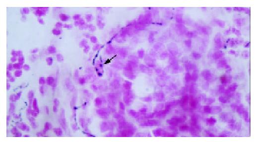
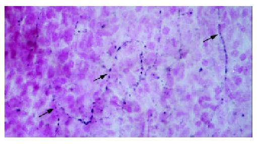
Figure 2 L-ENK-immunoreactive nerve fibers in esophageal carcinoma.
The fine nerve fiber (arrow) with varicosities on it encircled a tumor cell and contacted with it closely ×500.
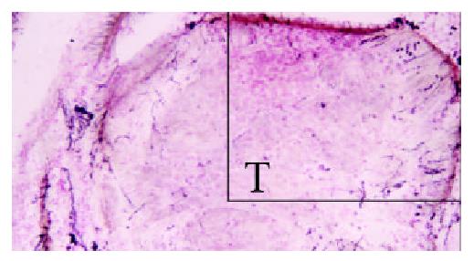
Figure 3 GAL-immunostaining of esophageal carcinoma.
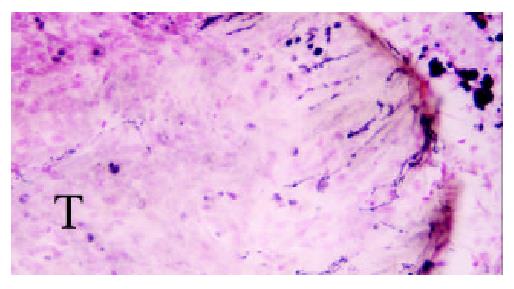
Many nerve fibers entered into a nestlike cancerous tissue (T) convergently from collective tissue around it. The structure on the top right corner was amplified in Figure 4×125.
Figure 4 Higher magnification of the part on the top right in Fig.
3. GAL- immunoreactive nerve fibers entered into esoph-ageal carcinoma from peripheral areas × 250.
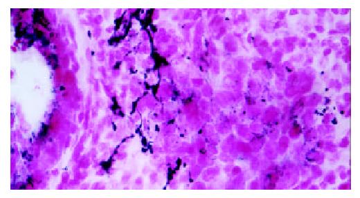
Figure 5 Section of Cardiac adenocarcinoma immunostained for GAL.
Many immunoreactive nerve fibers with beads-like varicosities distributed in the parenchyma of tumor ×500.
Figure 6 GAL-immunoreactive nerve fibers extended into nestlike cancerous tissue formed by cardiac adenocarcinoma cells.
The scattered nerve fibers contacted with tumor cells closely ×500.
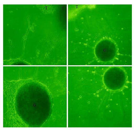
Figure 7 Montage photographs showing the effects of esoph-ageal (a) and cardiac (b) carcinoma tissue block (T) on co-cul-tured DRG (G).
On the side adjacent to tumor, DRG extending long and dense processes whereas on the opposite side, the pro-cesses are sparse and short (b), or no processes at all (a) ×125.
- Citation: Lü SH, Zhou Y, Que HP, Liu SJ. Peptidergic innervation of human esophageal and cardiac carcinoma. World J Gastroenterol 2003; 9(3): 399-403
- URL: https://www.wjgnet.com/1007-9327/full/v9/i3/399.htm
- DOI: https://dx.doi.org/10.3748/wjg.v9.i3.399


