Copyright
©The Author(s) 2003.
World J Gastroenterol. Oct 15, 2003; 9(10): 2216-2220
Published online Oct 15, 2003. doi: 10.3748/wjg.v9.i10.2216
Published online Oct 15, 2003. doi: 10.3748/wjg.v9.i10.2216
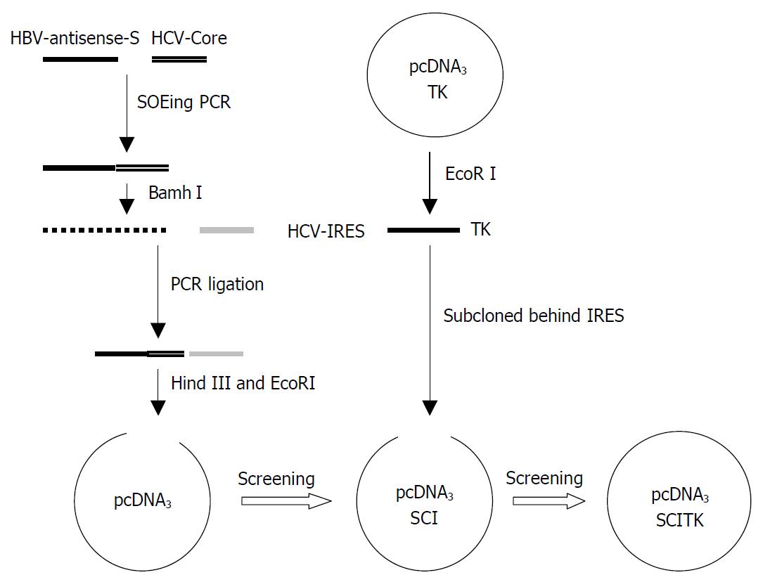
Figure 1 Construction scheme of recombinant plasmid pcDNA3-SCITK.
Figure 2 A: RT-PCR products of HCV IRES element.
M: marker (2 kb), Lanes 1-3: HCV IRES element (330 bp). B. Component segment analysis of pcDNA3-SCITK by agarose gel electrophoresis. M: marker (2 kb), Lanes 1-2: SCI-TK fragment demonstrated by PCR with HBV-S gene sense primer and TK antisense primer (5’-ACTTCCGTGGCTTCTTGCTG-3’ (nt 150-170). Lane 3: SCI fragment verified by PCR with HBV-S gene sense primer and HCV IRES antisense primer. Lane 4: SI segment from the ligation of HBV-S and HCV-Core gene by SOEing PCR. Lane 5: RT-PCR product of HCV-Core gene. Lane 6: Partial antisense segment of HBV-S gene.
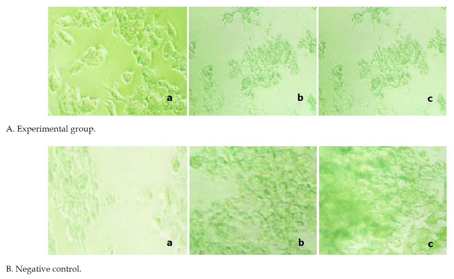
Figure 3 Effects of pcDNA3-SCITK expression on morphological alterations of 2.
2.15 and HepG2 cells. Photographs a, b and c in each group exhibited respectively the cell changes on the 1st, 3rd and 6th day post transfection. Lethiferous changes of 2.2.15 cells transfected with pcDNA3-SCITK were noted initially on the 3rd day of posttransfection and aggravated on the 6th day after transfection, in which the majority of cells were observed shrunken, rounded in shape and even dead. However, the negative contrl HepG2 cells grew well.
-
Citation: Kan QC, Yu ZJ, Lei YC, Hao LJ, Yang DL. Lethiferous effects of a recombinant vector carrying thymidine kinase suicide gene on 2.2.15 cells
via a self-modulating mechanism. World J Gastroenterol 2003; 9(10): 2216-2220 - URL: https://www.wjgnet.com/1007-9327/full/v9/i10/2216.htm
- DOI: https://dx.doi.org/10.3748/wjg.v9.i10.2216


