Copyright
©The Author(s) 2003.
World J Gastroenterol. Oct 15, 2003; 9(10): 2169-2173
Published online Oct 15, 2003. doi: 10.3748/wjg.v9.i10.2169
Published online Oct 15, 2003. doi: 10.3748/wjg.v9.i10.2169
Figure 1 Pre-operative CT scan shows the tumor originating in caudate lobe, post-opera-tive CT scan shows splitting line (arrow).
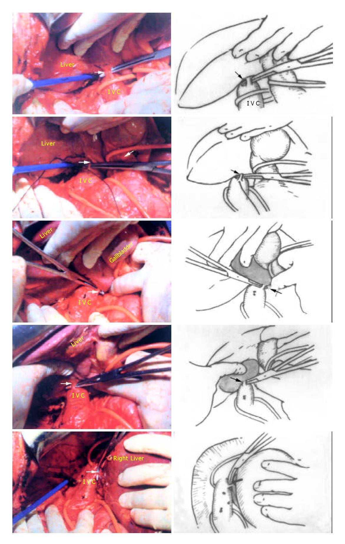
Figure 2 Five short hepatic veins ligated from caudal direction to cranial direction respestively (Arrow).
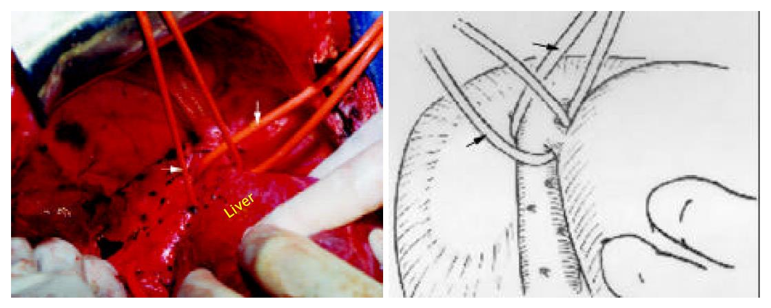
Figure 3 The suprahepatic inferior vena cava and right hepatic vein (RHV) dissected and encircled with tapes (thick arrow: RHV, thin arrow: suprahepatic IVC).
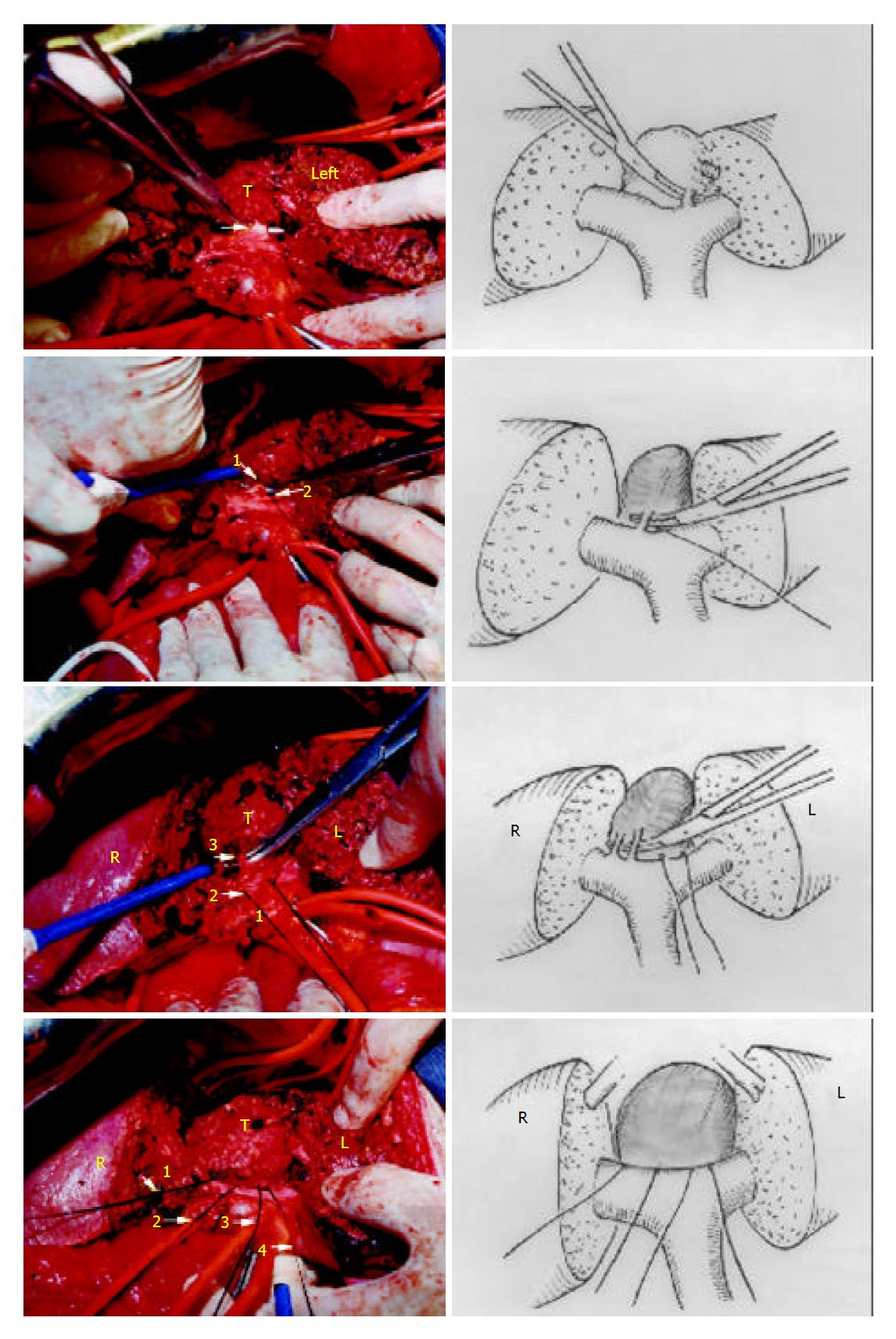
Figure 4 Four groups of portal triads to the caudate lobe(Arrow) were divided, the tumor was detached from the hilum.
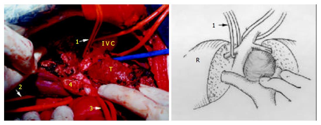
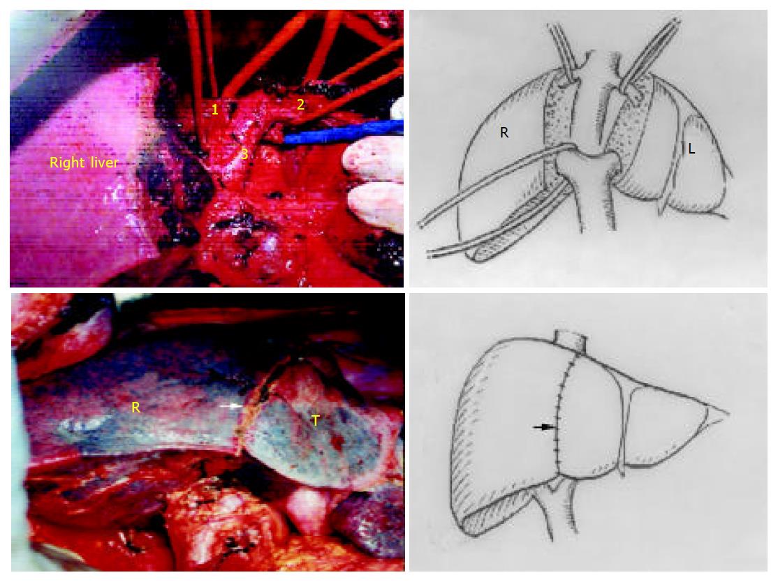
Figure 5 The tumor still attaches to MHV (1: tape across RHV, 2: tape across IVC, 3: tape across pedicle, 4: the portion of bifurcation, T: tumor).
Figure 6 Completely resected tumor.
Two halves of the liver were sutured. (1: RHV, 2: common trunk of MHV and LHV, 3: IVC, R: right liver, L: left liver, Arrow: interlobar plane, T: tumor).
- Citation: Peng SY, Li JT, Mou YP, Liu YB, Wu YL, Fang HQ, Cao LP, Chen L, Cai XJ, Peng CH. Different approaches to caudate lobectomy with “curettage and aspiration” technique using a special instrument PMOD: A Report of 76 cases. World J Gastroenterol 2003; 9(10): 2169-2173
- URL: https://www.wjgnet.com/1007-9327/full/v9/i10/2169.htm
- DOI: https://dx.doi.org/10.3748/wjg.v9.i10.2169


