Copyright
©The Author(s) 2003.
World J Gastroenterol. Oct 15, 2003; 9(10): 2154-2159
Published online Oct 15, 2003. doi: 10.3748/wjg.v9.i10.2154
Published online Oct 15, 2003. doi: 10.3748/wjg.v9.i10.2154
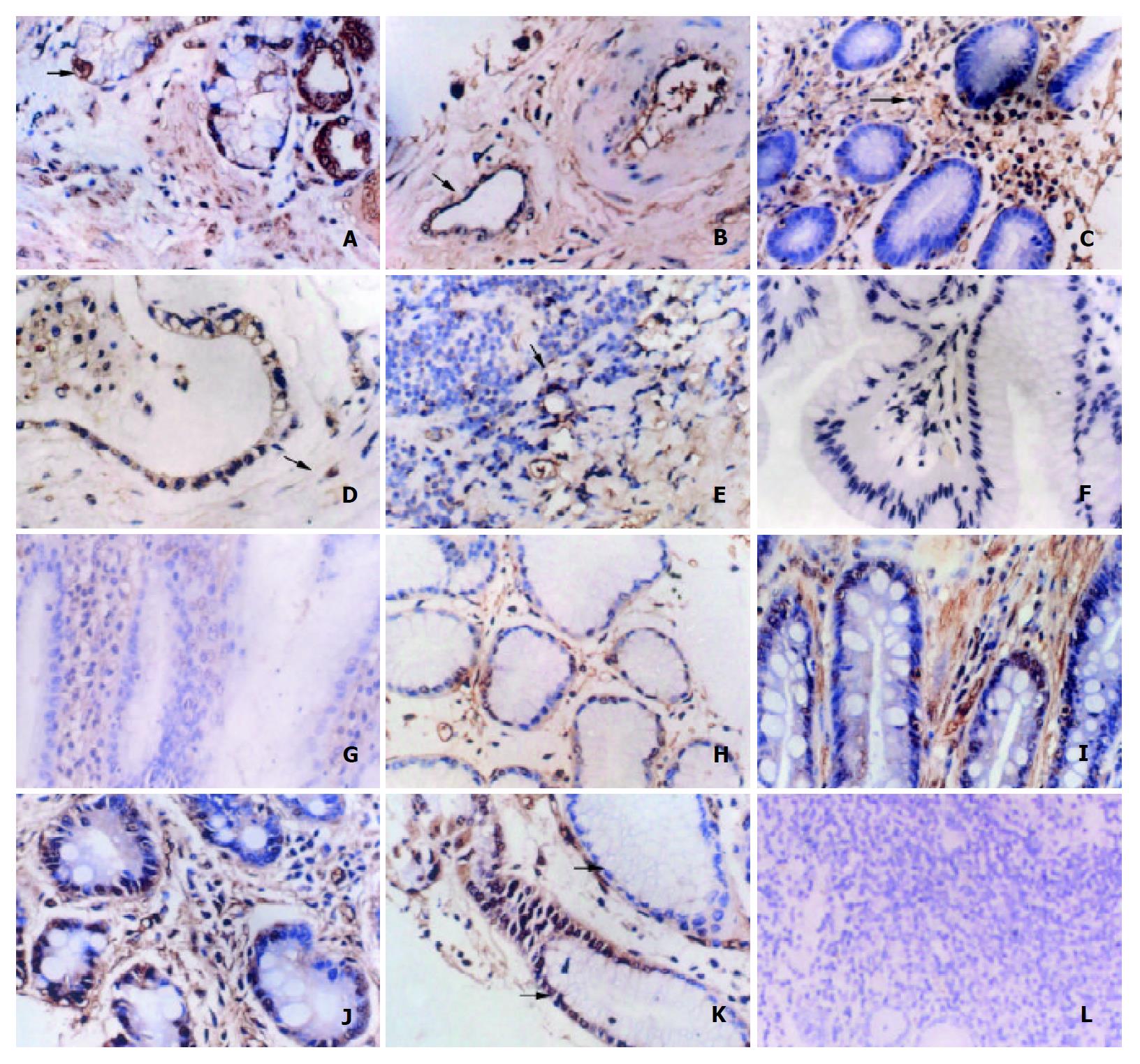
Figure 1 A: Ets1 immunohistochemical stain in gastric cancer cells.
The arrowed cell is a typical Ets1 positively stained cell. The area of stain was mainly, located in the nucleus of epithelial tumor cells. Ets1 protein was localized in both cytoplasm and nucleus of the stromal cells. B: Ets1 expression in vascular endothelial cells. This was an example of pooly differentiated gastric adenocarcinoma. Ets1 was present in endothelial cells of blood vessels (arrowed). C: Ets1 is expressed in interstitial cells. Photomi-crograph of a typical gastric carcinoma. Ets1 positive stromal cells were detected in the interstitial tissue. D: The expression of Ets1 in polarity. Image of gastric cancer cells infiltrating to the muscular layer. Intense staining of Ets1 was observed at the junction of the infiltrating tumor cells and the serosa. We termed it the polarity expression. E: Ets1 expression in breast carcinoma. Example of breast carcinoma, Ets1 positive cells arrowed. F: Ets1 negative control staining. G: Ets1 is not expressed in glandular cells of benign gastric ulcer. H-J: Ets1 expression in tissues with different histopathological grades. Section of grade I (H), grade II (I) and grade III (J) gastric cancer showing increased positive cells and intensity of Ets1 staining with advanced disease. K: Ets1 expression is high in undifferentiated tumor cell. The image contained well differentiated and poorly differentiated tumor tissues. The num-ber of positive cells and the intensity of the Ets1 staining were increased in undifferentiated tumor cells. L: Ets1 is not expressed in GIST. Photomicrograph of gastrointestinal stromal tumor (GIST). No Ets1 positive cancer cells were observed. (A, H, I, J, K origi-nal magnification × 400), (B, C, D, E, F, G original magnification × 100), (L, original magnification × 50).
- Citation: Yu Y, Zhang YC, Zhang WZ, Shen LS, Hertzog P, Wilson TJ, Xu DK. Ets1 as a marker of malignant potential in gastric carcinoma. World J Gastroenterol 2003; 9(10): 2154-2159
- URL: https://www.wjgnet.com/1007-9327/full/v9/i10/2154.htm
- DOI: https://dx.doi.org/10.3748/wjg.v9.i10.2154


