Copyright
©The Author(s) 2003.
World J Gastroenterol. Jan 15, 2003; 9(1): 141-147
Published online Jan 15, 2003. doi: 10.3748/wjg.v9.i1.141
Published online Jan 15, 2003. doi: 10.3748/wjg.v9.i1.141
Figure 1 Morphological characteristics of liver graft-derived DCs propagated for 4 d in the presence of GM-CSF.
A: DC from whole liver graft 4 d after transplantation (× 1200 b). B: DC from partial liver graft 4 d after transplantation (× 600b).
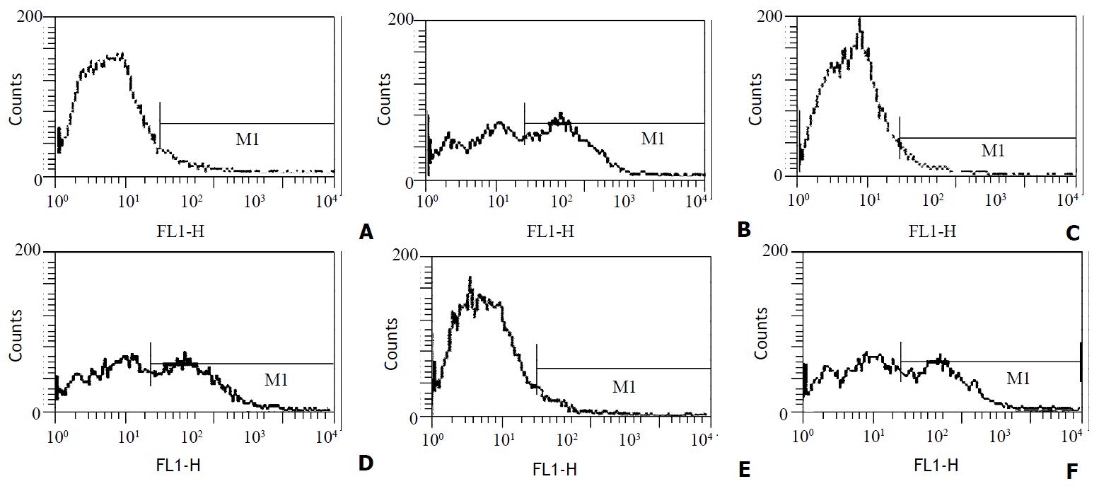
Figure 2 Expression of costimulatory molecules in DCs from liver grafts 4 d after transplantation by FCM.
A: Expression of CD40 in whole liver graft-derived DCs; B: Expression of CD40 in partial liver graft-derived DCs; C: Expression of CD80 in whole liver graft-derived DCs; D: Expression of CD80 in partial liver graft-derived DCs; E: Expression of CD86 in whole liver graft-derived DCs; F: Expression of CD86 in partial liver graft-derived DCs.
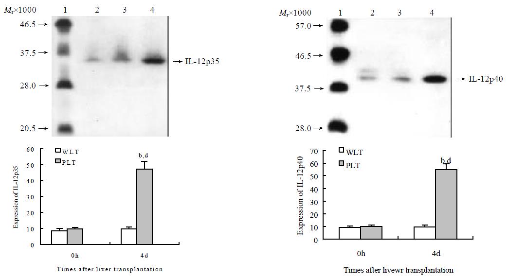
Figure 3 Detection of IL-12 proteins in liver graft-derived DCs by Western blotting.
Lanes 1: protein marker. Lanes 2-4: extracts derived from DCs from liver graft 0 h after transplantation, whole liver graft (WLT) and partial liver graft (PLT) 4 d after transplantation, respectively. Compared with 4 d WLT group, bP < 0.001; Compared with 0 h PLT group, dP < 0.001.
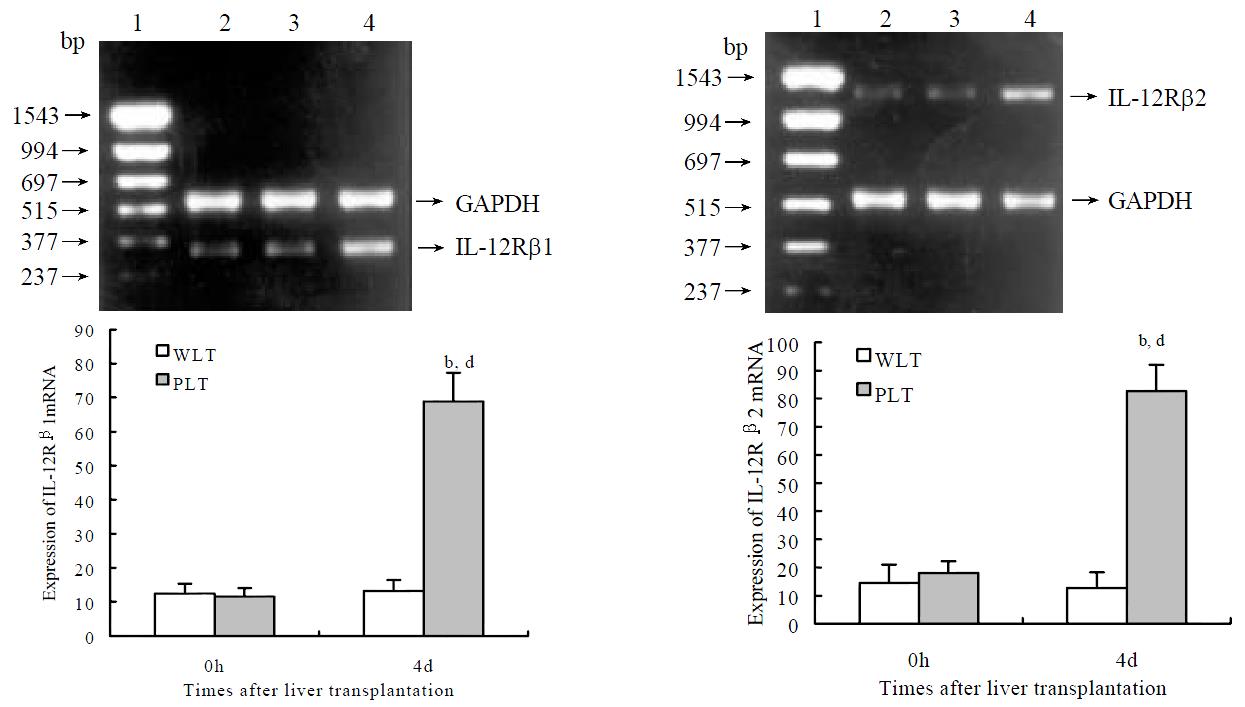
Figure 4 Detection of IL-12 R mRNA in liver graft-derived DCs by RT-PCR.
Lanes 1: marker; Lanes 2-4: extracts derived from DCs from liver graft 0 h after transplantation, whole liver graft (WLT) and partial liver graft (PLT) 4 d after transplantation, respectively. Compared with 4 d WLT group, bP < 0.001; Compared with 0 h PLT group, dP < 0.001.
- Citation: Xu MQ, Yao ZX. Functional changes of dendritic cells derived from allogeneic partial liver graft undergoing acute rejection in rats. World J Gastroenterol 2003; 9(1): 141-147
- URL: https://www.wjgnet.com/1007-9327/full/v9/i1/141.htm
- DOI: https://dx.doi.org/10.3748/wjg.v9.i1.141


