Copyright
©The Author(s) 2002.
World J Gastroenterol. Dec 15, 2002; 8(6): 1129-1133
Published online Dec 15, 2002. doi: 10.3748/wjg.v8.i6.1129
Published online Dec 15, 2002. doi: 10.3748/wjg.v8.i6.1129
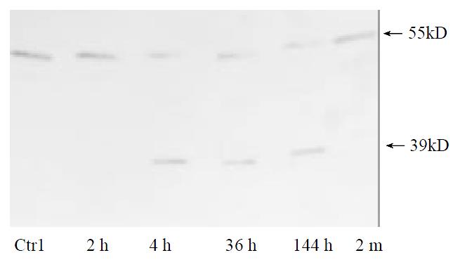
Figure 1 TNFR1 shedding on the surface of parenchymal hepatocytes occurs in regenerative liver.
Data shown represent time course of TNFR1 shedding after partial hepatectomy. Plasma membrane from normal liver was used as control.
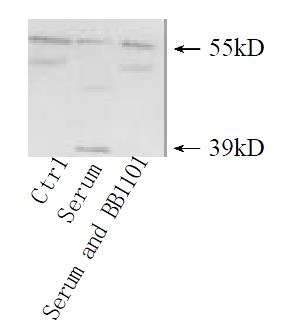
Figure 2 TNFR1 shedding under the stimulation of serum from rats after partial hepatectomy.
Cultured hepatocytes were treated with serum from regener-ative liver at 36 h for 30 min, or the serum accompanied with metalloprotase inhibitor BB1101 at 2 mmol·L-1. Cultured hepatocytes under no any treatments were used as control.
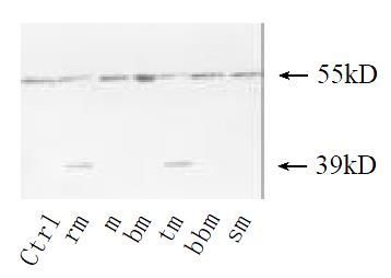
Figure 3 TNFR1 shedding induced with plasma membrane of hepatocytes from regenerative liver.
Cultured hepatocytes were treated for 30 min with plasma membrane from hepatocytes at 36 h after hepatectomy (rm) or sham-operated (m) or from hepa-tocytes treated with TNFα at 10 µg·mL-1 for 2 h (tm). Plasma membrane boiled for 5 min (bm) as control. Then metalloprotase inhibitor BB1101 at 2 mmol·L-1 (bbm), or staurosporine at 5 ng·mL-1 e (sm) was added into culture medium. Cultured hepa-tocytes under no any treatments were as control.
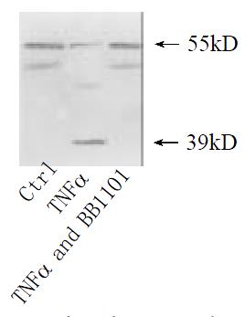
Figure 4 TNFR1 shedding under the stimulation of TNF α.
Cultured hepatocytes were treated respectively with TNFα at 10 μg·mL-1 for 2 h, or TNFα accompanied with metalloprotase inhibitor BB1101 at 2 mmol·L-1. Cultured hepatocytes under no any treatments were as control.
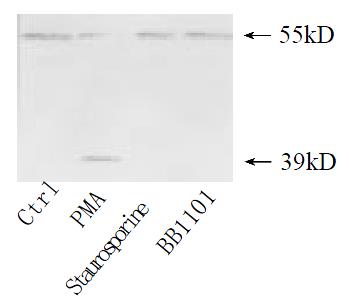
Figure 5 Effect of PMA on TNFR1 shedding.
Cultured hepa-tocytes were treated with PMA at 10 µg·mL-1 for 30 min, staurosporine accompanied with PMA at 5 ng·mL-1, or PMA accompanied with BB1101 at 2 mmol·L-1. Confluent cultured hepatocytes without any treatment were used as control.
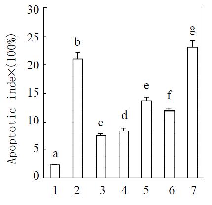
Figure 6 Induction of apoptosis after treatment with TNFα.
Means of data shown in figure were: 1. Control; 2. Treated with TNFα at 10 μg·mL-1; 3. Treatment with TNFα at 10 μg·mL-1 after treated with plasma membrane purified from liver at 36 h after partial hepatectomy at 2 μg·mL-1; 4. Treated with plasma membrane at 2 μg·mL-1 purified from hepatocytes induced with TNFa accompanied with TNFa at 10 μg·mL-1; 5. Treated with PMA at a concentration of 10 μmol·L-1 (in DMSO) accompa-nied with TNF at 10 μg·mL-1; 6. Treated with serum from rat after partial hepatectomy for 36 h at 5% accompanied with TNFα at 10 μg·mL-1; 7. Treated with TNFα at 10 μg·mL-1 after treated with plasma membrane purified from rat liver regen-erated for 2 months at 2 μg·mL-1. Apoptotic index = (numbers of apoptotic cells/total cells numbers per well) × 100. Data were means from 6 separate experiments × SE (n = 6 wells). Differ-ent letters over bars indicate significant differences, P < 0.05. The results are confirmed by DNA fragmentation by agarose electrophoresis (data not provided).
- Citation: Xia M, Xue SB, Xu CS. Shedding of TNFR1 in regenerative liver can be induced with TNF α and PMA. World J Gastroenterol 2002; 8(6): 1129-1133
- URL: https://www.wjgnet.com/1007-9327/full/v8/i6/1129.htm
- DOI: https://dx.doi.org/10.3748/wjg.v8.i6.1129


