Copyright
©The Author(s) 2002.
World J Gastroenterol. Dec 15, 2002; 8(6): 1067-1072
Published online Dec 15, 2002. doi: 10.3748/wjg.v8.i6.1067
Published online Dec 15, 2002. doi: 10.3748/wjg.v8.i6.1067
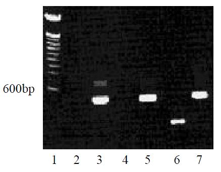
Figure 1 MCP-3 mRNA expression of CMT93 or its gene transfectants (RT-PCR).
Lane 1: 100bp DNA ladder marker; Lane 2:CMT93+Primer MCP-3; Lane 3: CMT93 + Primer β-actin; Lane 4: CMT93/Mock + Primer MCP-3; Lane 5: CMT93/Mock + Primer β-actin; Lane 6: CMT93/MCP-3 + Primer MCP-3; Lane 7: CMT93/MCP-3 + Primer β-actin.
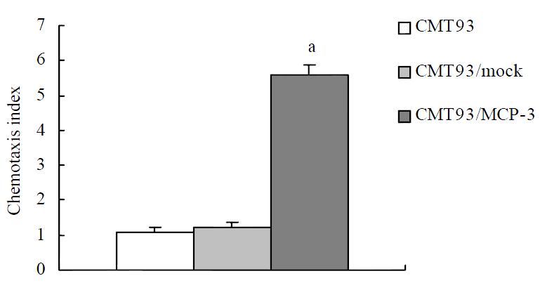
Figure 2 Chemotactic activity of MCP-3 in the supernatant from CMT93 cells or its gene transfectants.
P < 0.05 vs CMT93 or CMT93/mock group.
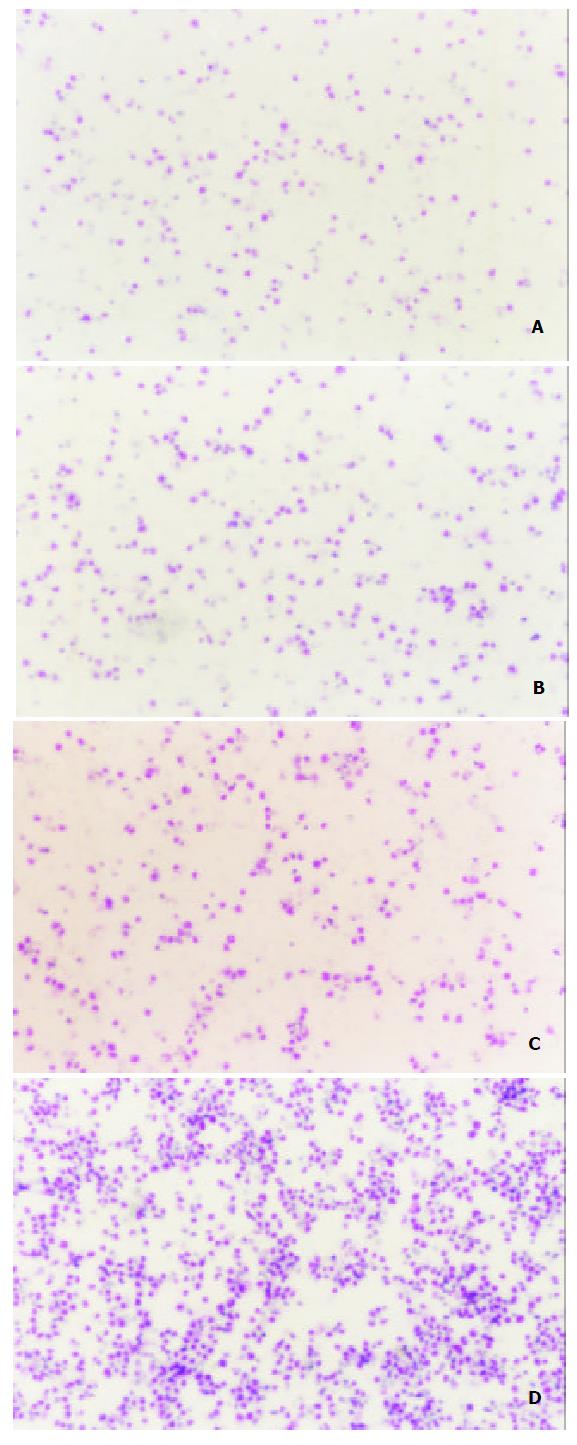
Figure 3 Chemotactic activity of MCP-3 in supernatant from CMT93 or its transfectants (Wright staining showed the chemoattracted target cells × 25).
A: medium; B: CMT93 supernatant. The chemoattracted cells were not increased significantly; C: CMT93/Mock supernatant. The chemoattracted cells were not increased significantly; D: CMT93/MCP-3 supernatant. The chemoattracted cells were increased significantly.
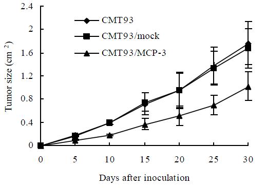
Figure 4 The tumor growth curves of CMT93 or its gene transfectants
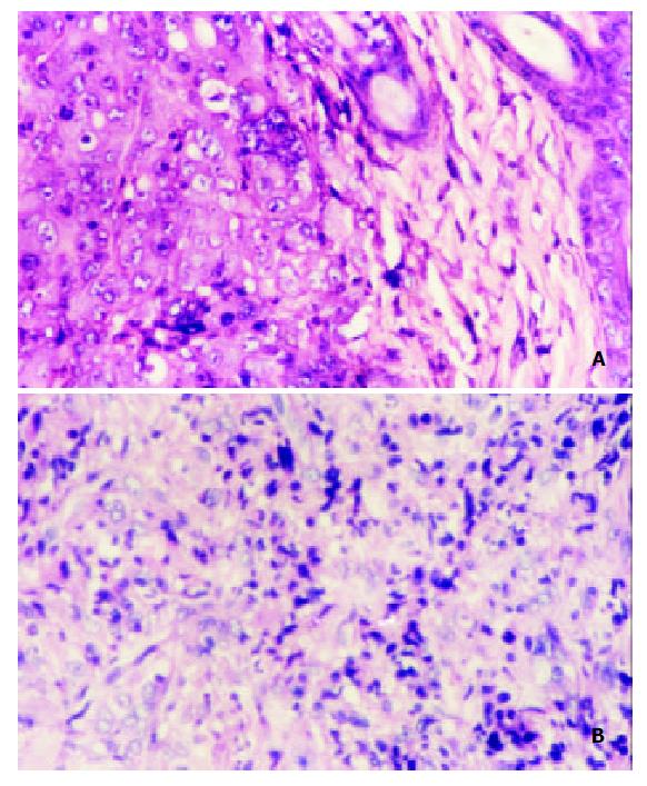
Figure 5 Histopathology of the tumors derived from CMT93 or CMT93/MCP3 (HE staining × 200).
A: tumor derived from CMT93 cells; few infiltrated immune cells were found; B:tu-mor derived from CMT93/MCP-3 cells, the infiltrated immune cells were increased.
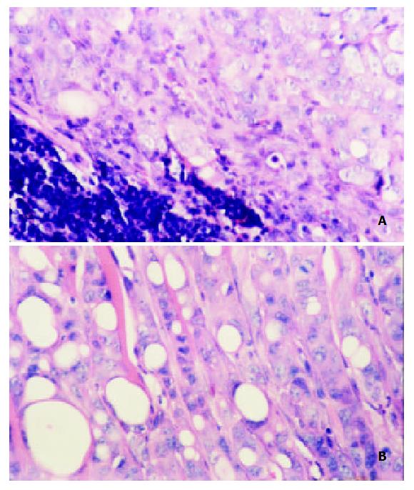
Figure 6 Histopathology of tumor derived from CMT93 me-tastasized to the local drainage lymph node(HE staining × 200).
A: up-left is the lymph tissue,down-right is the tumor tissue; B: tumor infiltrated to the muscle tissue.
- Citation: Hu JY, Li GC, Wang WM, Zhu JG, Li YF, Zhou GH, Sun QB. Transfection of colorectal cancer cells with chemokine MCP-3 (monocyte chemotactic protein-3) gene retards tumor growth and inhibits tumor metastasis. World J Gastroenterol 2002; 8(6): 1067-1072
- URL: https://www.wjgnet.com/1007-9327/full/v8/i6/1067.htm
- DOI: https://dx.doi.org/10.3748/wjg.v8.i6.1067


