Copyright
©The Author(s) 2001.
World J Gastroenterol. Dec 15, 2001; 7(6): 880-883
Published online Dec 15, 2001. doi: 10.3748/wjg.v7.i6.880
Published online Dec 15, 2001. doi: 10.3748/wjg.v7.i6.880
Figure 1 CTA of portal vein that show tributary of splenic vein clearly.
Figure 2 indirect portography via transsplenic artery show splenic vein and its tributary.
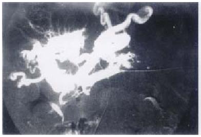
Figure 3 direct portography show main portal vein obstruction and the varices.
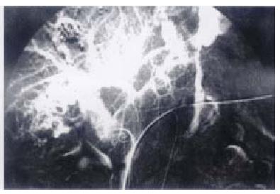
Figure 4 the same patient as figure 3, the variceal blood flow was turned slowly after embolization.
- Citation: Gong GQ, Wang XL, Wang JH, Yan ZP, Cheng JM, Qian S, Chen Y. Percutaneous transsplenic embolization of esophageal and gastrio-fundal varices in 18 patients. World J Gastroenterol 2001; 7(6): 880-883
- URL: https://www.wjgnet.com/1007-9327/full/v7/i6/880.htm
- DOI: https://dx.doi.org/10.3748/wjg.v7.i6.880


