Copyright
©The Author(s) 2001.
World J Gastroenterol. Aug 15, 2001; 7(4): 547-550
Published online Aug 15, 2001. doi: 10.3748/wjg.v7.i4.547
Published online Aug 15, 2001. doi: 10.3748/wjg.v7.i4.547
Figure 1 Dynamic changes of serum ALT, AST and HGV-RNA in patient Hu.
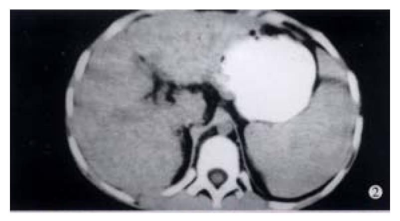
Figure 2 CT picture displays uneven liver density, small nodular changes and splenomegaly extended in an area of eight costae.
EM × 15000
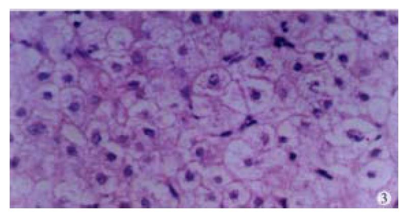
Figure 3 Histological changes of the liver showing cloudy swelling of hepatocytes.
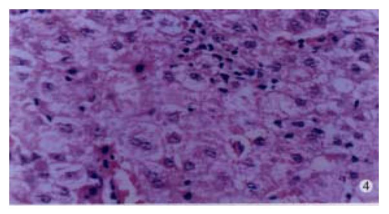
Figure 4 Histological changes of the liver in a patient with acute single-infected hepatitis G showing punctate necrosis, focal lymphocyte infiltration, and acidophilic degeneration of hepatocytes.
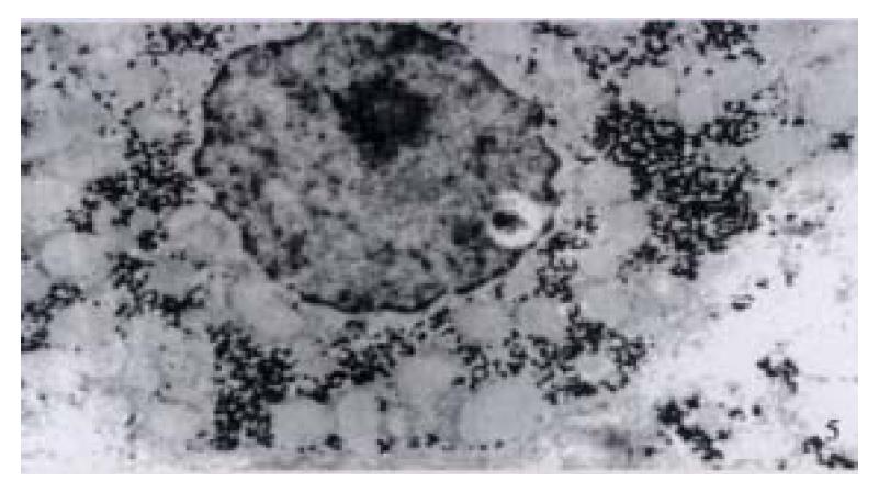
Figure 5 Ultrastructure of the liver tissues in a patient with acute simple HGV infection showing shirinkage of liver cells with zigzag edges, extension of rough surfaced endoplastic reticulum of hepatocytes, proliferation of collagen fibrils extended into the cytoplasm of hepatocytes.
EM × 15000
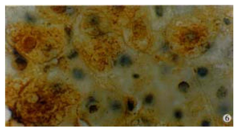
Figure 6 Immunohistochemical preperation stained by specific HGV McAb for HGVNS5 in a patient with single HG infection showing brown-yellow granules presented mostly in cytoplasm of hepatocytes, and partially in the nuclei.
DAB staining, hematoxylin staining, BA staining. (oil) × 1000
- Citation: Xu JZ, Yang ZG, Le MZ, Wang MR, He CL, Sui YH. A study on pathogenicity of hepatitis G virus. World J Gastroenterol 2001; 7(4): 547-550
- URL: https://www.wjgnet.com/1007-9327/full/v7/i4/547.htm
- DOI: https://dx.doi.org/10.3748/wjg.v7.i4.547


