Copyright
©The Author(s) 2001.
World J Gastroenterol. Aug 15, 2001; 7(4): 510-514
Published online Aug 15, 2001. doi: 10.3748/wjg.v7.i4.510
Published online Aug 15, 2001. doi: 10.3748/wjg.v7.i4.510
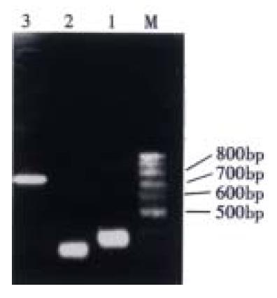
Figure 1 RT-PCR of VH, VL and ScFv fragment of MG7 antibody.
1: VH; 2: VL; 3: ScFv; M: 100 bp ladder
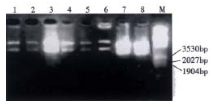
Figure 2 Enzymatic analysis of MG7 recombinant phage antibody library with Eco RI and Hin dIII.
1-8: Recombinant clones from library; M: λ/Eco RI and Hin dIII
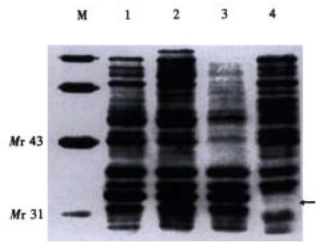
Figure 3 Measurement of the relative molecular weight of soluble MG7 ScFv.
1-3: Periplasmic extracts; 4: Neg. ctrl; M: Low molecular mass protein marker
-
Citation: Yu ZC, Ding J, Nie YZ, Fan DM, Zhang XY. Preparation of single chain variable fragment of MG
7 mAb by phage display technology. World J Gastroenterol 2001; 7(4): 510-514 - URL: https://www.wjgnet.com/1007-9327/full/v7/i4/510.htm
- DOI: https://dx.doi.org/10.3748/wjg.v7.i4.510


