Copyright
©The Author(s) 2001.
World J Gastroenterol. Aug 15, 2001; 7(4): 500-505
Published online Aug 15, 2001. doi: 10.3748/wjg.v7.i4.500
Published online Aug 15, 2001. doi: 10.3748/wjg.v7.i4.500
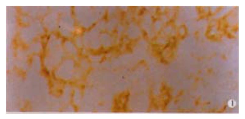
Figure 1 Immunohistochemical staining for VEGF in adenocarcinoma tissues of the stomach.
The positive signal was found mainly on the surface and in the cytoplasm of gastric cancer cells. × 400
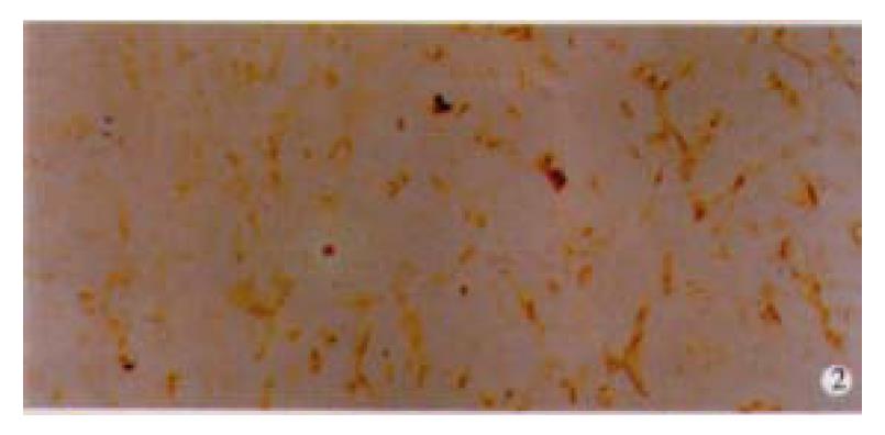
Figure 2 Immunohistochemical staining for flk-1/KDR in undifferentiated cancer tissues of the stomach.
The positive signal was found on the surface of endothelial cells. × 100
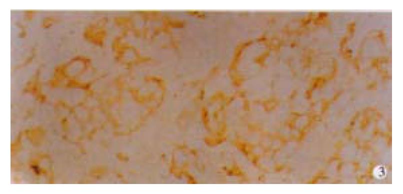
Figure 3 Immunohistochemical staining for flk-1/KDR in adenocarcinoma tissues of the stomach.
It was found mainly on the surface and in the cytoplasm of gastric cancer cells. × 200
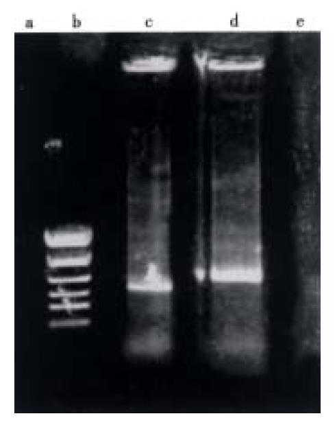
Figure 4 PCR analysis of SGC-s-hVEGF and SGC-as-hVEGF.
a. SGC-s-hVEGF/SP6+b; b. PCR marker; c. SGC-s-hVEGF/SP6+a; d. SGC-as-hVEGF/SP6b; e. SGC-as-hVEGF/SP6+a
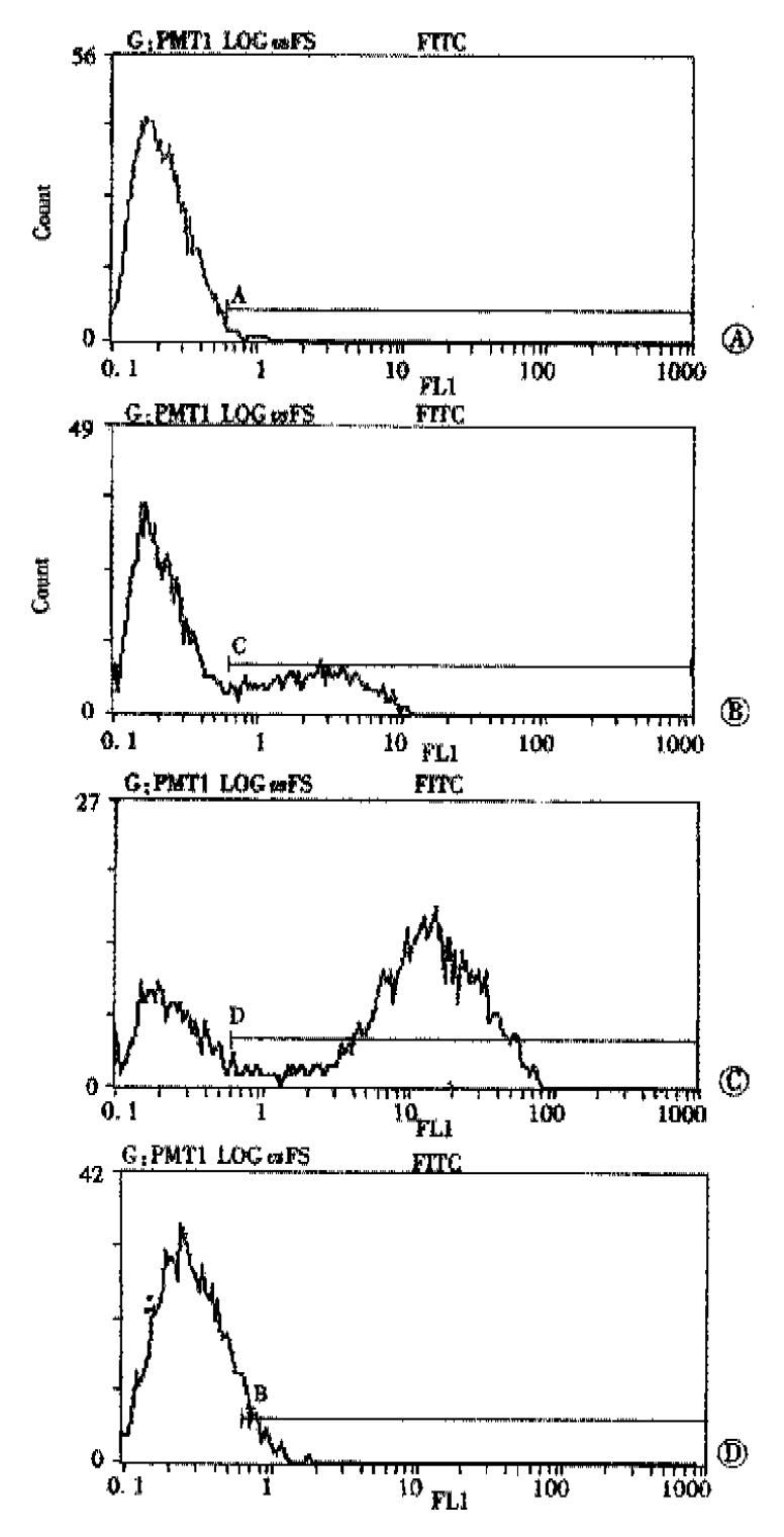
Figure 5 Changes in cell surface VEGF protein as deter mined by flow cytometer immunofluorescence staining analysis.
(A) Flow cytometric detection of VEGF changes of control. (B) Flow cytometric detection of VEGF changes of SGC-7901. (C) Flow cytometric detection of VEGF changes of SGC/s-hVEGF. (D) Flow cytometric detection of VEGF changes of SGC/as-hVEGF.
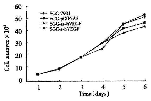
Figure 6 The growth curve of different transfectant.
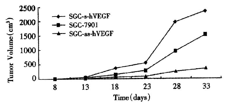
Figure 7 The volume of tumors in nude mice (n = 5/group) s.
c. developing from s.c. injection totaling 1.4 × 10⁶ cells/ animal of the different transfectants.
- Citation: Liu DH, Zhang XY, Fan DM, Huang YX, Zhang JS, Huang WQ, Zhang YQ, Huang QS, Ma WY, Chai YB, Jin M. Expression of vascular endothelial growth factor and its role in oncogenesis of human gastric carcinoma. World J Gastroenterol 2001; 7(4): 500-505
- URL: https://www.wjgnet.com/1007-9327/full/v7/i4/500.htm
- DOI: https://dx.doi.org/10.3748/wjg.v7.i4.500


