Copyright
©The Author(s) 2000.
World J Gastroenterol. Aug 15, 2000; 6(4): 532-539
Published online Aug 15, 2000. doi: 10.3748/wjg.v6.i4.532
Published online Aug 15, 2000. doi: 10.3748/wjg.v6.i4.532
Math 1 Math(A1).
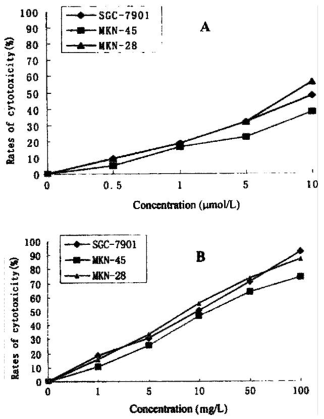
Figure 1 Cytotoxicity of As2O3 (A), and HCPT (B), on gastric cancer cells.
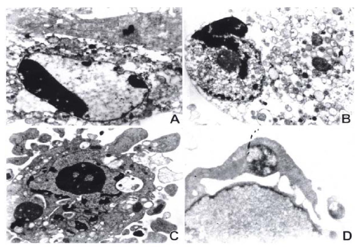
Figure 2 Electron microscopic observation of ultrastructural changes in gastric cancer cells treated with 10 μmol/L As2O3 for 48 h.
The early change of apoptosis, nuclear chromatin condensation, looks like a new moon subjacent to the nuclear membranes (A). The intermediate stage of apoptosis presents cytoplasmic blebs, nuclear condensation, overflow of nuclear chromatin, and like-sprout (B). The late stage of apoptosis is characterized by splitting of nuclear membrane, nuclear chr omatin condensation, overflow of some parts of nuclear chromatin, and fragmentation of nucleus (C), and formation of apoptotic bodies (C) (D).
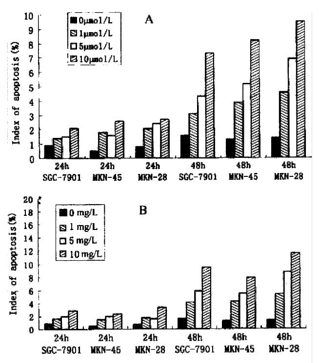
Figure 3 The index of apoptosis of gastric cancer cells induced by As2O3 (A) and by HCPT (B).
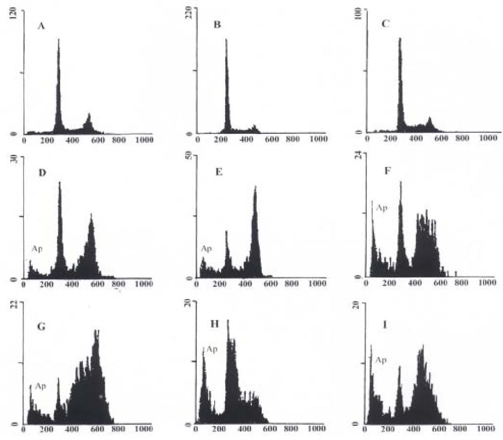
Figure 4 Nuclear DNA contents measured by flow cytometry in As2O3 and HCPT-induced apoptosis in gastric cancer cells at 48 h.
Ap represents apoptotic cells. A: Untreated MKN-45 cells; B: Untreated SGC-7901 cells; C: Untreate d MKN-28 cells; D: MKN-45 cells treated with 10 μmol/L As2O3; E: SGC-79 01 cells treated with 10 μmol/L As2O3; F: MKN-28 cells treated with 10 μmol/L As2O3; G: MKN-45 cells treated with 10 mg/L HCPT; H: SGC-7901 cells treated with 10 mg/L HCPT; I: MKN-28 cells treated with 10 mg/L HCPT.
- Citation: Tu SP, Zhong J, Tan JH, Jiang XH, Qiao MM, Wu YX, Jiang SH. Induction of apoptosis by arsenic trioxide and hydroxy camptothecin in gastriccancer cells in vitro. World J Gastroenterol 2000; 6(4): 532-539
- URL: https://www.wjgnet.com/1007-9327/full/v6/i4/532.htm
- DOI: https://dx.doi.org/10.3748/wjg.v6.i4.532


