Copyright
©The Author(s) 1999.
World J Gastroenterol. Oct 15, 1999; 5(5): 424-429
Published online Oct 15, 1999. doi: 10.3748/wjg.v5.i5.424
Published online Oct 15, 1999. doi: 10.3748/wjg.v5.i5.424
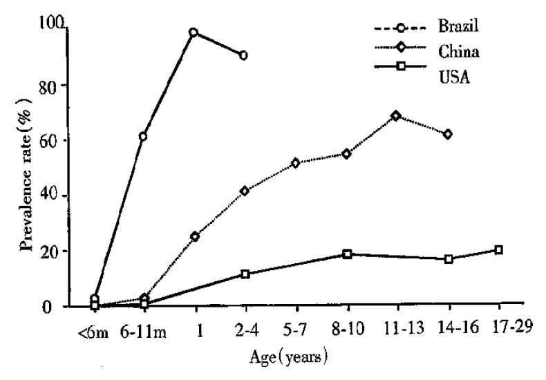
Figure 1 Prevalence of IgG antibodies to C.
parvum in different populations from Brazil, China and the United States. M = month. Data reproduced from reference 3 with perission.
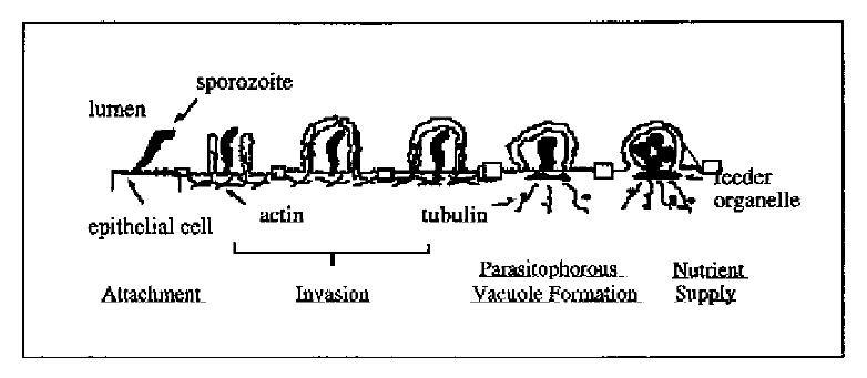
Figure 2 Molecular model for C.
parvum infection of epithelial cells. C. parvum sporozoite attaches specifically to the apical membrane surface of epithelial cells. The attachment induces rearrangement of the host cell cytoskeleton and facilitates the parasite’s invasion and parasitophorous vacuole formation. Host cell cytoskeleton rearrangement forms the feeder organelle underlying the parasite attachment site and facilitates the uptake of nutrients by the parasite from the host cell cytoplasm.
- Citation: Chen XM, LaRusso NF. Human intestinal and biliary cryptosporidiosis. World J Gastroenterol 1999; 5(5): 424-429
- URL: https://www.wjgnet.com/1007-9327/full/v5/i5/424.htm
- DOI: https://dx.doi.org/10.3748/wjg.v5.i5.424


