Copyright
©The Author(s) 1999.
World J Gastroenterol. Oct 15, 1999; 5(5): 397-403
Published online Oct 15, 1999. doi: 10.3748/wjg.v5.i5.397
Published online Oct 15, 1999. doi: 10.3748/wjg.v5.i5.397
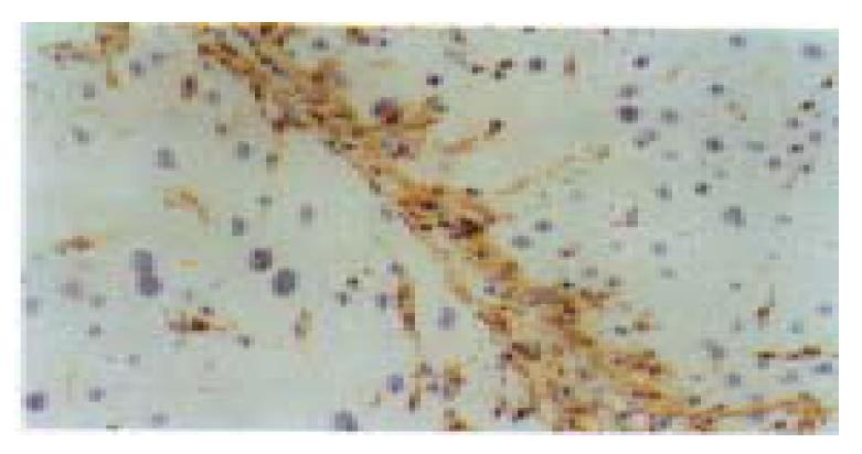
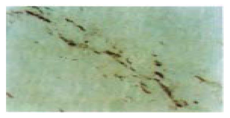
Figure 1 The cells in the cytofibrotic septa were mainly composed of desmin (Dm) positive hepatic stellate cells (HSCs) and myo fibroblasts (MFs) and some Dm negative fibroblasts (Fbs) (8 weeks of CCl4 administration).
Immunohistochemical staining for Dm, × 200
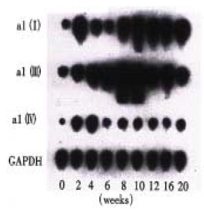
Figure 2 Dynamic changes of α1(I), α1(III) and α1(IV) procollagen mRNA expression in different stages of the experiment (Northern blot analysis).
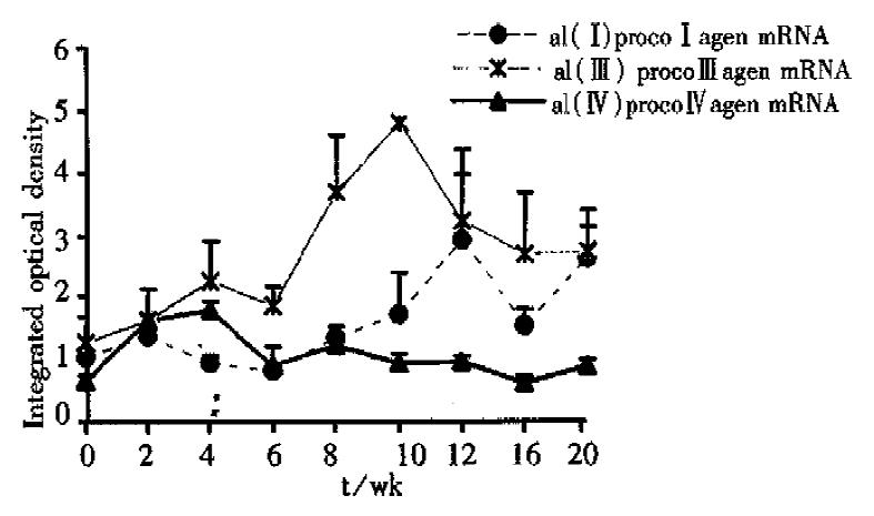
Figure 3 The changes of α1(I), α1(III) and α1(IV) procollagen mRNA expression in different stages of the experiment (Northern blot analysis).
Figure 4 Most of the cells in the cytofibrotic septa expressed α1(III)- procollagen mRNA (6 weeks of CCl4 administration).
In situ-hibridization, × 400
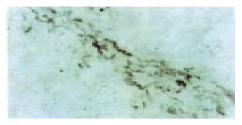
Figure 5 The serial tissue section of Figure 4, only a few of the cells in the cytofibrotic septa expressed α1(I) procollagen mRNA.
In situ hybridization, × 400
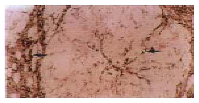
Figure 6 Some spindle cells and the endothelial cell s of small blood (arrow) vessels in the fibrotic septa and the endothelial cells of the capillized sinusoids (arrow) expressed strong signals of á1 (IV) procollagen mRNA in the late stage of the experiment (16 weeks of CCl4 administration).
In situ hybridization, × 200
- Citation: Du WD, Zhang YE, Zhai WR, Zhou XM. Dynamic changes of type I, III and IV collagen synthesis and distribution of collagen-producing cells in carbon tetrachloride-induced rat liver fibrosis. World J Gastroenterol 1999; 5(5): 397-403
- URL: https://www.wjgnet.com/1007-9327/full/v5/i5/397.htm
- DOI: https://dx.doi.org/10.3748/wjg.v5.i5.397


