Copyright
©The Author(s) 1999.
World J Gastroenterol. Aug 15, 1999; 5(4): 345-348
Published online Aug 15, 1999. doi: 10.3748/wjg.v5.i4.345
Published online Aug 15, 1999. doi: 10.3748/wjg.v5.i4.345
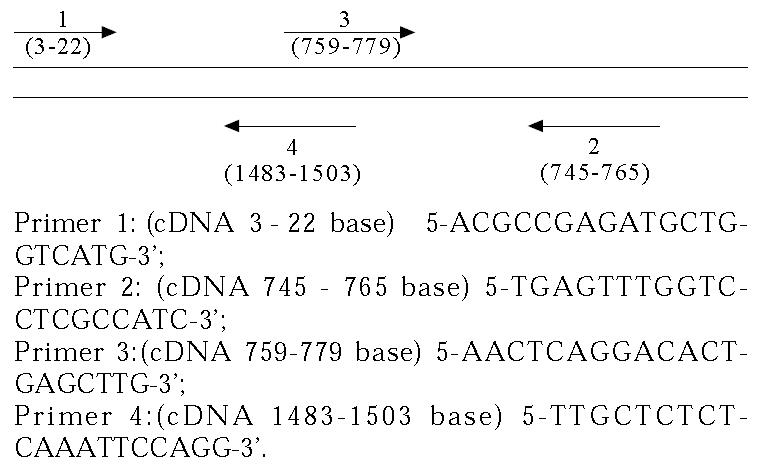
Math 1 Math(A1).
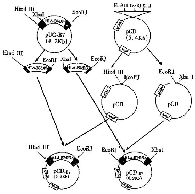
Figure 1 Construction procedure of eukaryotic expression vector.
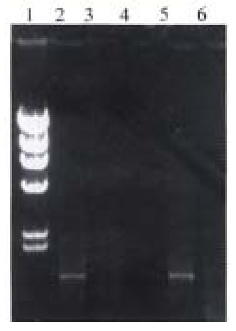
Figure 2 The electrophoresis results of RT-PCR of HLA-B7 cDNA.
1. λDNA/Hind III marker; 2. The full-length of cDNA (1.5 kb); 3. The upstream region of cDNA (750 bp); 6. The downstream region of cDNA (750 bp)
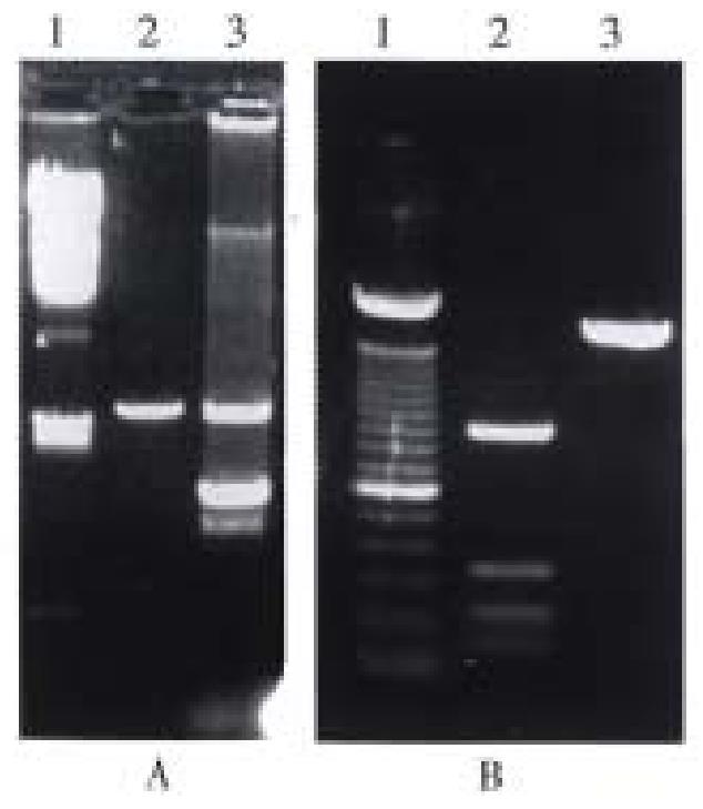
Figure 3 The results of restriction analysis of pUC-B7.
A. 1. λDNA/Hind III marker; 2. pUC19/Hind III; 3. pUC-B7/Hind I + EcoR I; B. 1. pGEM/Hinf I + Rsa I + Sin I marker; 2. cDNA/Hinf I; 3. cDNA (1.5 kb)
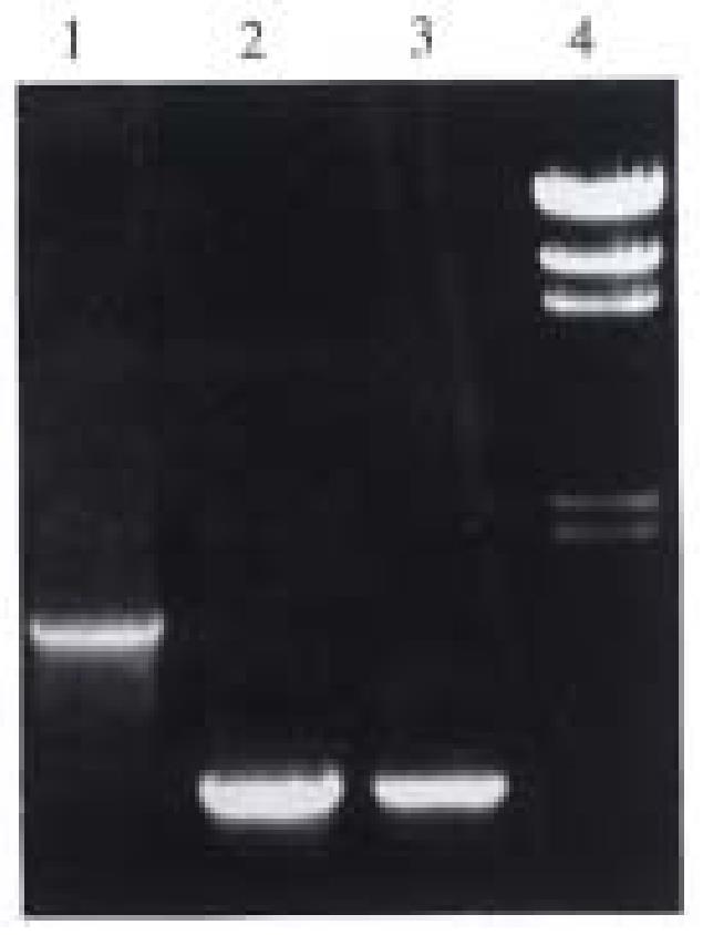
Figure 4 The results of PCR detection of pUC-B7; 1.
Full length of cDNA (1.5 kb); 2. The upstream region of cDNA (750 bp); 3. The downstream region of cDNA (750 bp); 4. λDNA/Hind III marker
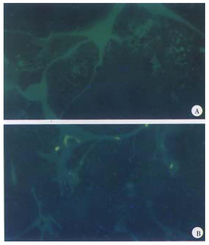
Figure 5 The expression of pCD-B7 in NIH/3T3 cells.
A. Negative control; B. Positive cells
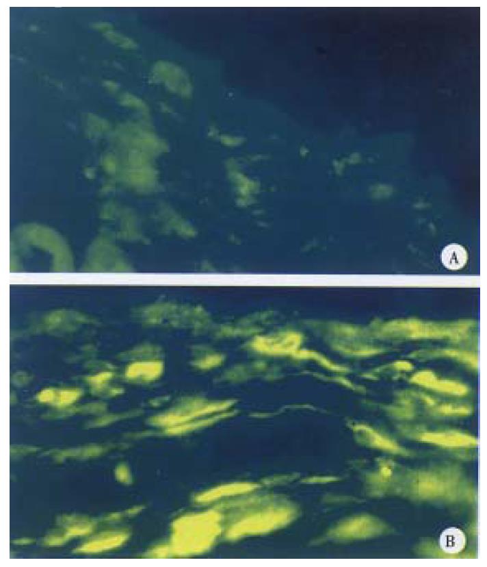
Figure 6 The results of pCD-B7 expressed in subcutaneous tissue of mice.
A. Negative control; B. Positive cells
- Citation: Shen Q, Li QF, Deng YZ, Zhang JM, Zhang J. Cloning and expression of HLA-B7 gene. World J Gastroenterol 1999; 5(4): 345-348
- URL: https://www.wjgnet.com/1007-9327/full/v5/i4/345.htm
- DOI: https://dx.doi.org/10.3748/wjg.v5.i4.345


