Copyright
©The Author(s) 1998.
World J Gastroenterol. Aug 15, 1998; 4(4): 303-306
Published online Aug 15, 1998. doi: 10.3748/wjg.v4.i4.303
Published online Aug 15, 1998. doi: 10.3748/wjg.v4.i4.303
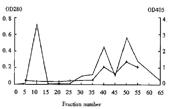
Figure 1 Chromatogram of digested Hb3.
OD280 (·-·) shows concentration of protein. OD405 (+ - +) shows immunoreactivity.
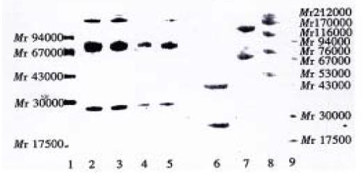
Figure 2 SDS-PAGE results of antibody electrophoresis.
Lane 1: low molecular weight marker; lane 2-3: intact Hb3; lane 4-5: intact Hb3 (1/10 concentration); lane 6: Hb3 fragments F(ab’)2 (broken); lane 7: Hb3 fragments F(ab’)2 (unbroken); lane 8: high molecular weight marker; lane 9: low molecular weight marker.
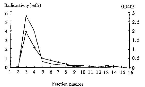
Figure 3 Chromatogram of 99mTc labeled antibody, OD405 shows immunoreactivity.
·-· radioactivity, + - + immunoreactivity.
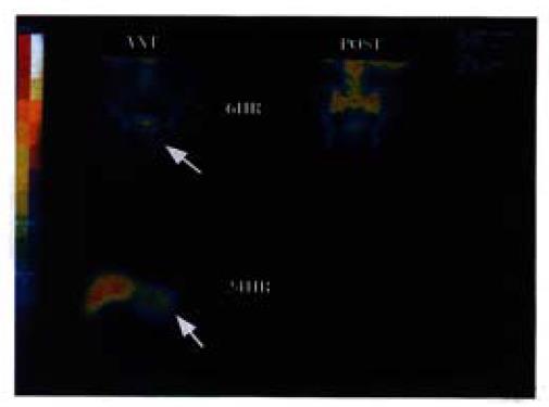
Figure 4 Images of Patient 4.
Rectal cancer complicated with liver metastasis. Anterior scan (6 h) shows an unclear abnormal radiation concentration under the bladder (upper left). Anterior scan (24 h) shows a clear abnormal radiation concentration in the left lobe of liver (lower left).
- Citation: Hu JY, Su JZ, Pi ZM, Zhu JG, Zhou GH, Sun QB. Radioimmunoimaging of colorectal cancer using 99mTc-labeled monoclonal antibody. World J Gastroenterol 1998; 4(4): 303-306
- URL: https://www.wjgnet.com/1007-9327/full/v4/i4/303.htm
- DOI: https://dx.doi.org/10.3748/wjg.v4.i4.303


