Copyright
©The Author(s) 1998.
World J Gastroenterol. Aug 15, 1998; 4(4): 287-293
Published online Aug 15, 1998. doi: 10.3748/wjg.v4.i4.287
Published online Aug 15, 1998. doi: 10.3748/wjg.v4.i4.287
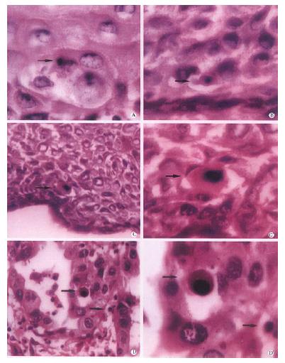
Figure 1 Apoptotic cells in human esophageal epithelia (H & E staining).
Micrographs illustrate the apoptotic cells in normal tissue (A) and the tissues with lesions of BCH (B), DYS (C) and SCC (D). × 400. C’ and D’ were the higher magnification of C and D. × 1000. Scattered apoptotic cells (arrow) were located among the compartments of dysplastic and hyperproliferatic cells. The apoptotic bodies most often appeared as a single structure separated from the surrounding intact cells by a clear halo (C).
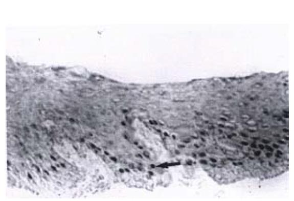
Figure 2 Immunostaining of Waf1p21 in biopsy samples of esophageal BCH.
The Waf1p21-positive immunostaining cells were located at the third and forth cell layers of the epithelium (arrow). × 400
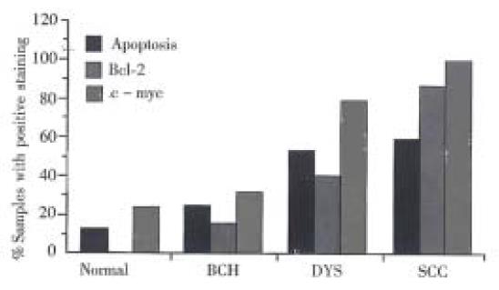
Figure 3 Changes of apoptosis and immunoreactivity of bcl-2 and c-myc in esophageal normal epithelia and epithelia with different severities of lesions.
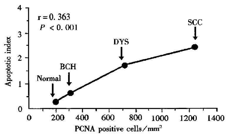
Figure 4 Changes of apoptosis and cell proliferation determined by PCNA in esophageal carcinogenesis.
- Citation: Wang LD, Zhou Q, Wei JP, Yang WC, Zhao X, Wang LX, Zou JX, Gao SS, Li YX, Yang C. Apoptosis and its relationship with cell proliferation, p53, Waf1p21, bcl-2 and c-myc in esophageal carcinogenesis studied with a high-risk population in northern China. World J Gastroenterol 1998; 4(4): 287-293
- URL: https://www.wjgnet.com/1007-9327/full/v4/i4/287.htm
- DOI: https://dx.doi.org/10.3748/wjg.v4.i4.287


