Copyright
©The Author(s) 2025.
World J Gastroenterol. Mar 7, 2025; 31(9): 100271
Published online Mar 7, 2025. doi: 10.3748/wjg.v31.i9.100271
Published online Mar 7, 2025. doi: 10.3748/wjg.v31.i9.100271
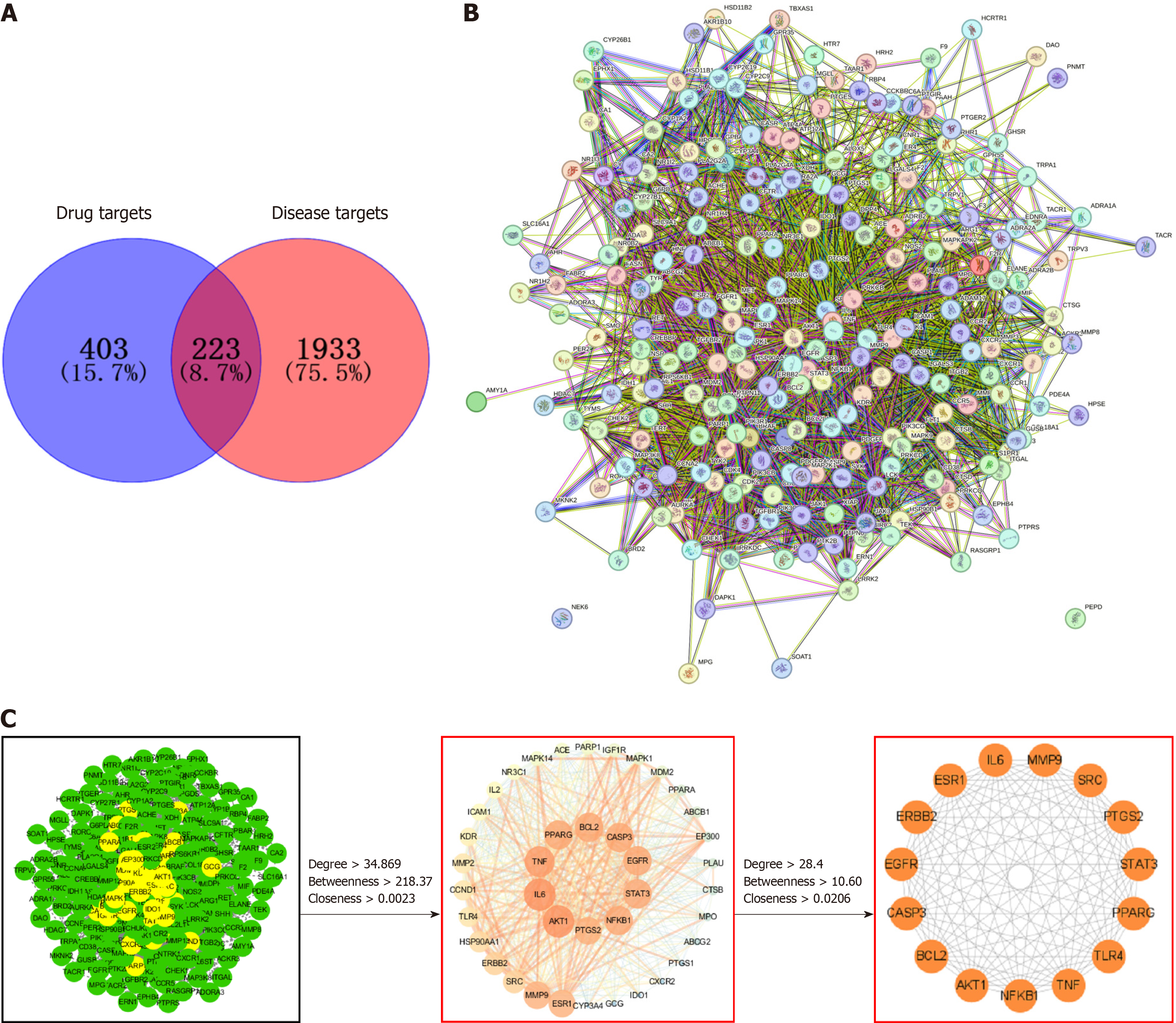
Figure 1 Protein-protein interaction network and topological analysis.
A: Venn diagram of the active ingredient of Panax ginseng (P. ginseng) and potential targets of ulcerative colitis (UC); B: Protein-protein interaction network acquired from the Search Tool for the Retrieval of Interacting Genes/Proteins database platform; C: Screening of hub genes for P. ginseng for the treatment of UC.
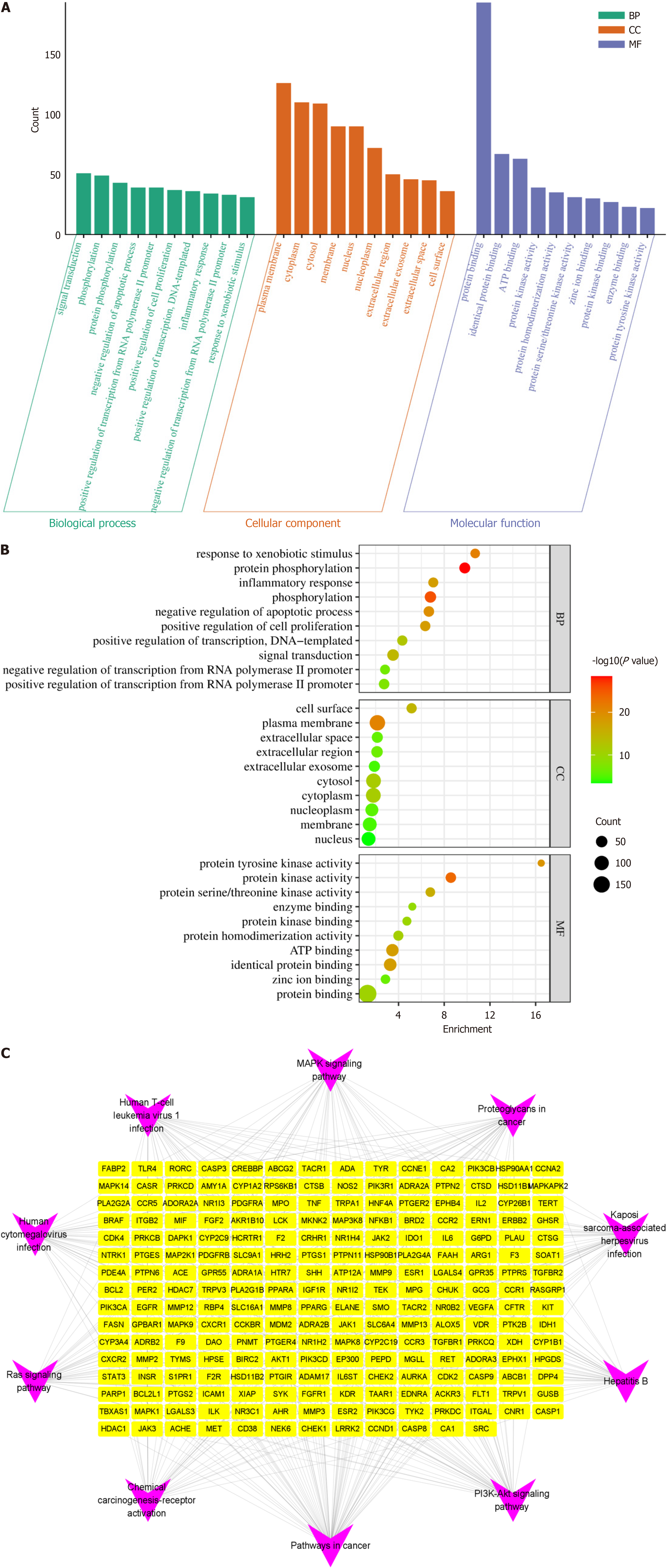
Figure 2 Gene Ontology and Kyoto Encyclopedia of Genes and Genomes enrichment analysis.
A: Histogram of Gene Ontology (GO) enrichment analysis, displaying categories for biological process, cellular component, and molecular function; B: Bubble chart of GO enrichment analysis, where the X-axis represents the enrichment value. The color intensity of the dots indicates the P value (with redder dots indicating lower P values), and the size of the dots reflects the number of counts; C: Network analysis of the Kyoto Encyclopedia of Genes and Genomes (KEGG) pathways. Purple nodes represent the top 10 KEGG pathways, and the yellow nodes represent the intersection genes between Panax ginseng and ulcerative colitis. BP: Biological process; CC: Cellular component; MF: Molecular function.
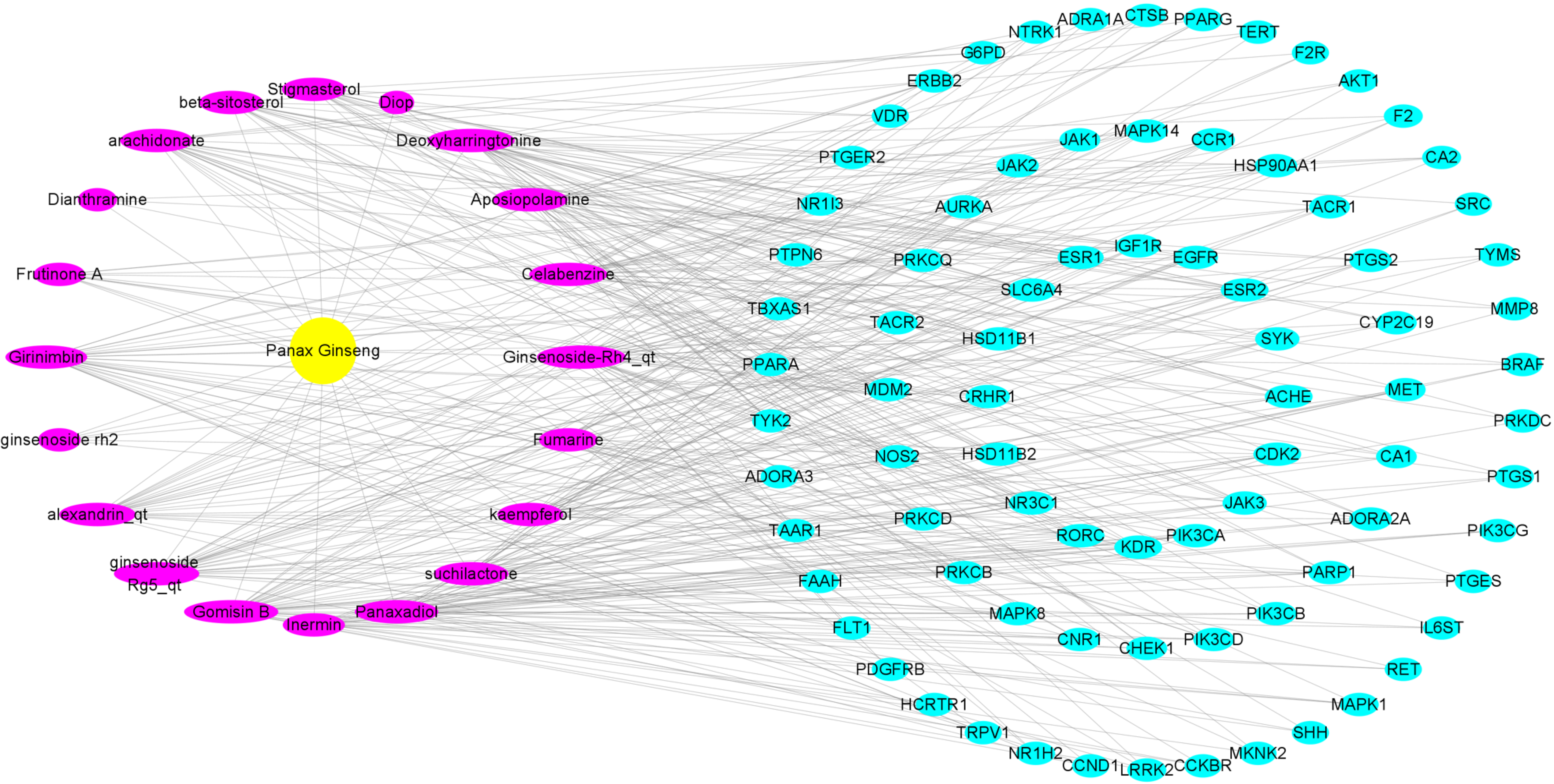
Figure 3 Screening of the main active ingredients of Panax ginseng for the treatment of ulcerative colitis.
Yellow nodes represent Panax ginseng, purple nodes represent its active ingredients, and cyan nodes represent intersection genes with a degree value > 2.
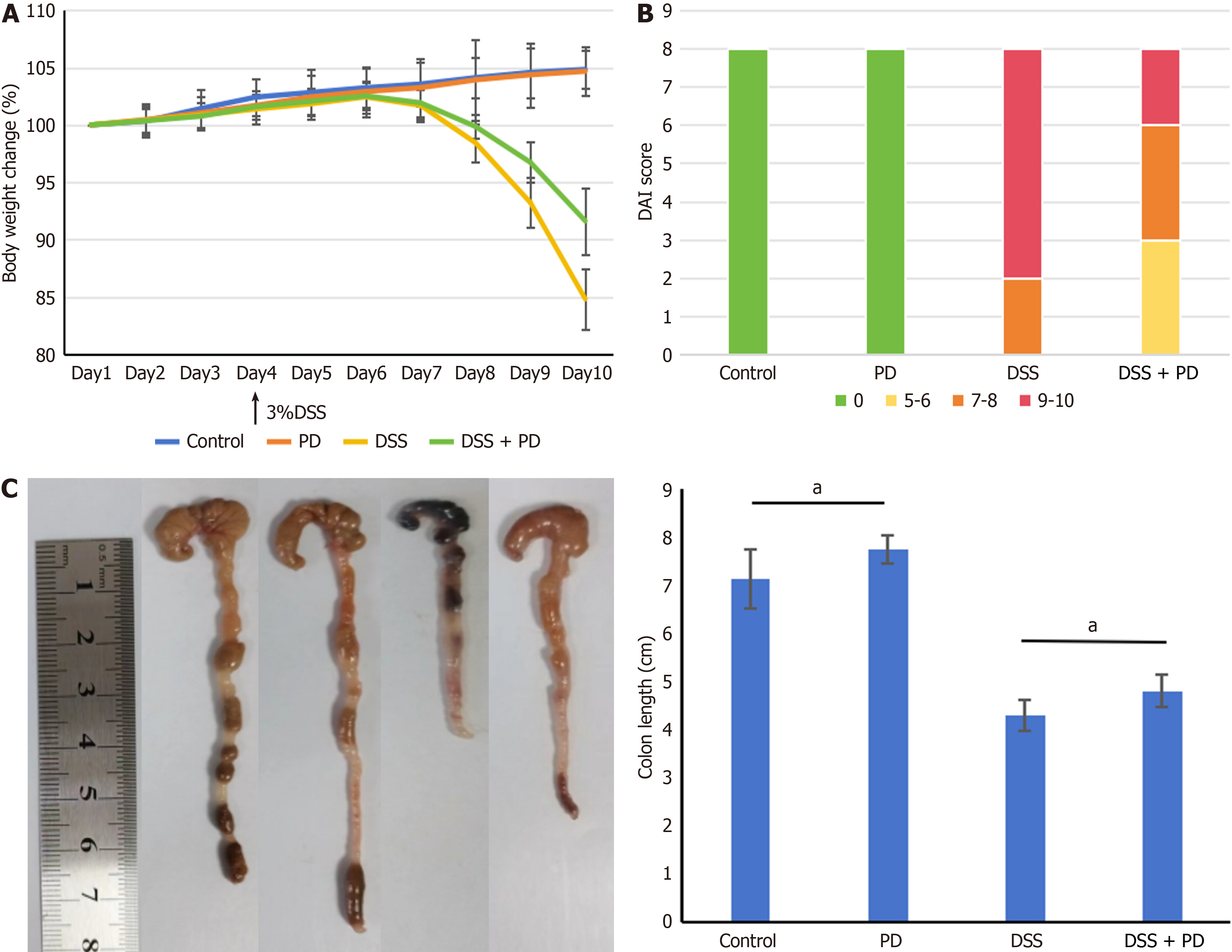
Figure 4 Panaxadiol alleviated weight loss, reduced the Disease Activity Index score, and prevented colon shortening in mice with dextran sulfate sodium-induced colitis.
A: Body weight changes; B: Disease Activity Index score; C: Colon length. Data are expressed as mean ± standard error of the mean (n = 8). aP < 0.05; DAI: Disease Activity Index; DSS: Dextran sulfate sodium; PD: Panaxadiol.
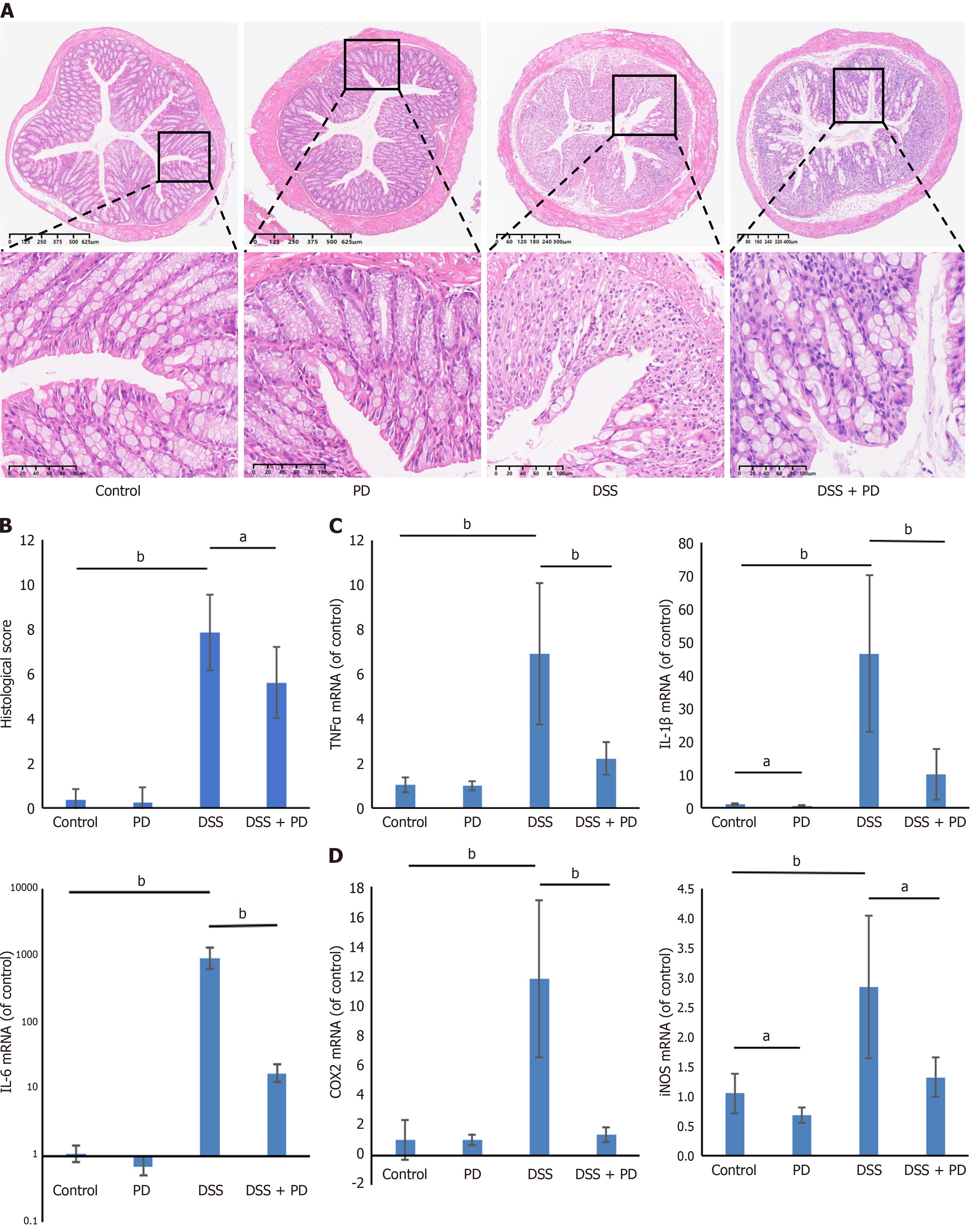
Figure 5 Panaxadiol attenuated intestinal inflammation in mice with dextran sulfate sodium-induced colitis.
A: Representative images of hematoxylin-eosin staining (× 40 and × 200 magnification); B: Histopathological score; C and D: Panaxadiol treatment reduces pro-inflammatory cytokines (C) and pro-inflammatory mediators (D) in mice with dextran sulfate sodium-induced colitis. Data are expressed as mean ± standard error of the mean (n = 6). aP < 0.05; bP < 0.01; DSS: Dextran sulfate sodium; PD: Panaxadiol.
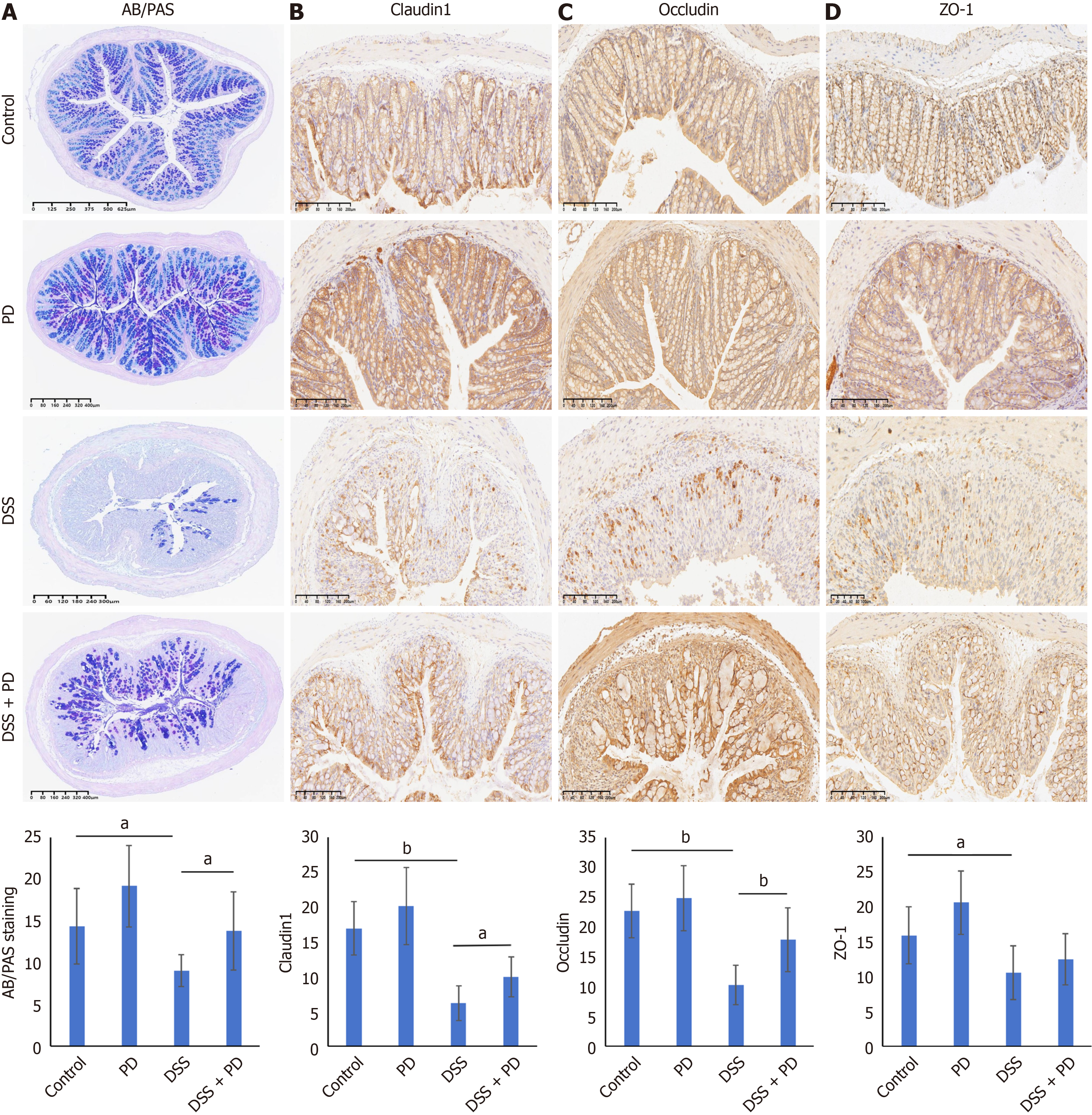
Figure 6 Panaxadiol maintained the integrity of the intestinal epithelial barrier in mice with dextran sulfate sodium-induced colitis.
A: Alcian blue and periodic acid-Schiff staining (× 40); B-D: Immunohistochemistry analyses of claudin-1, occludin, and ZO-1 (× 100 magnification). Data are expressed as mean ± standard error of the mean (n = 8). aP < 0.05; bP < 0.01; DSS: Dextran sulfate sodium; PD: Panaxadiol.
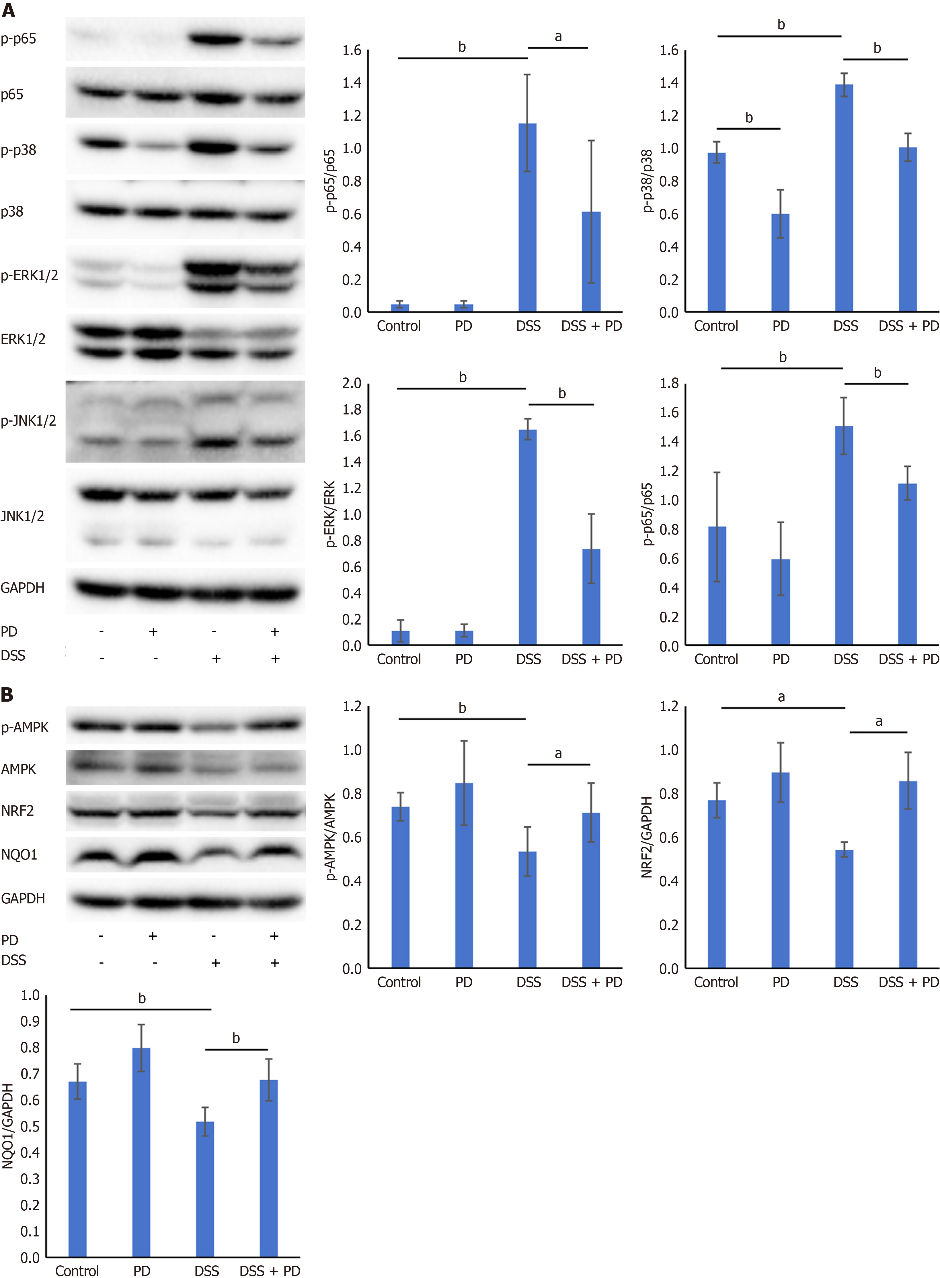
Figure 7 Panaxadiol regulated the activation of MAPK/NF-κB and AMPK/NRF2/NQO1 signaling in mice with dextran sulfate sodium-induced colitis.
A: Effect of panaxadiol on the MAPK/NF-κB signaling pathway; B: Effect of panaxadiol on the AMPK/NRF2/NQO1 signaling pathway. Data are expressed as mean ± standard error of the mean (n ≥ 3). aP < 0.05; bP < 0.01; DSS: Dextran sulfate sodium; PD: Panaxadiol.
- Citation: Qin Y, Zhang RY, Zhang Y, Zhao YQ, Hao HF, Wang JP. Network pharmacology and in vivo study: Unraveling the therapeutic mechanisms of Panax ginseng in potentially treating ulcerative colitis. World J Gastroenterol 2025; 31(9): 100271
- URL: https://www.wjgnet.com/1007-9327/full/v31/i9/100271.htm
- DOI: https://dx.doi.org/10.3748/wjg.v31.i9.100271


