Copyright
©The Author(s) 2025.
World J Gastroenterol. Feb 28, 2025; 31(8): 101585
Published online Feb 28, 2025. doi: 10.3748/wjg.v31.i8.101585
Published online Feb 28, 2025. doi: 10.3748/wjg.v31.i8.101585
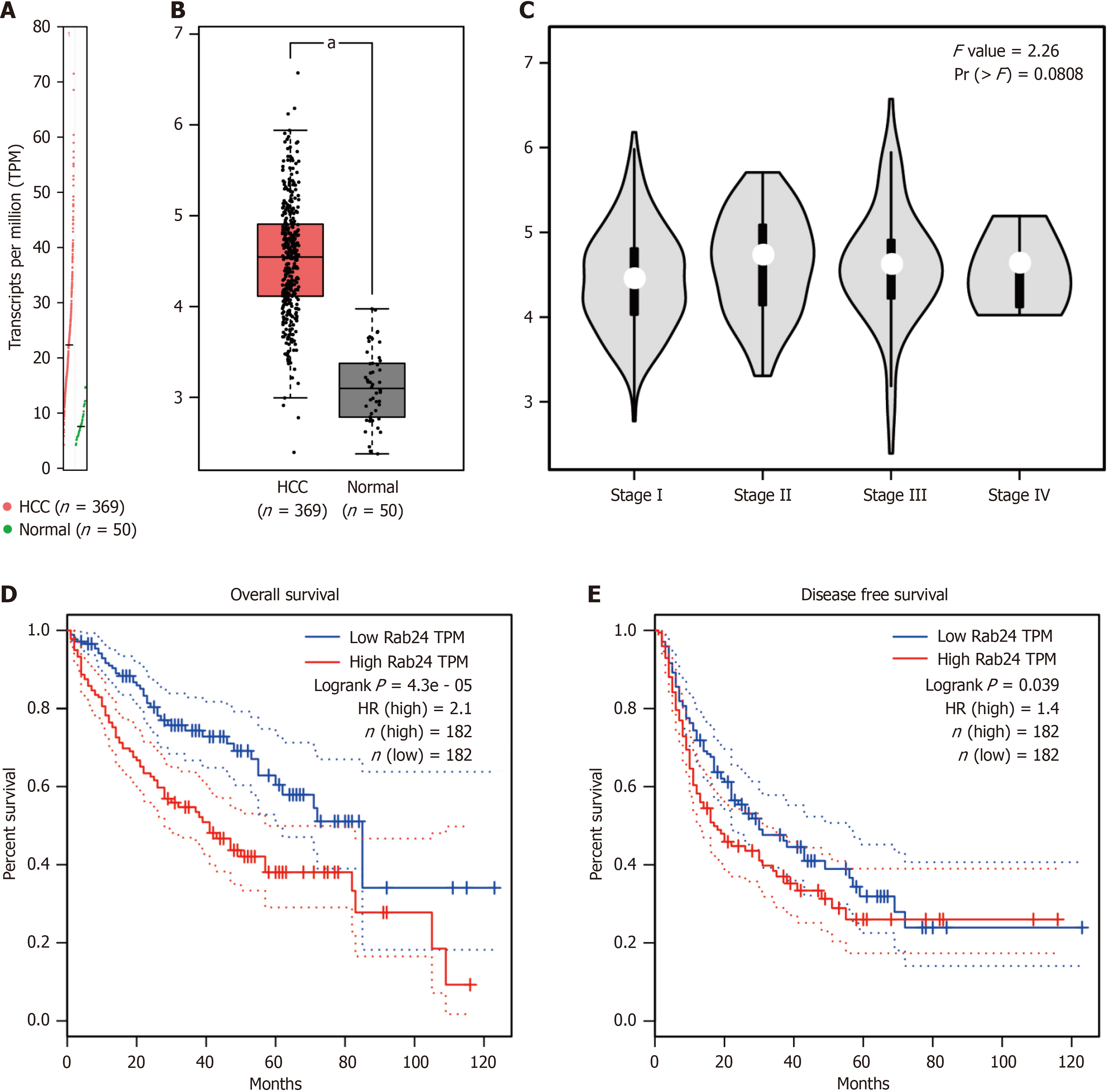
Figure 1 The transcription level of Rab24 in hepatocellular carcinoma and its prognostic role.
A: Scatterplot showed the transcriptional profile of Rab24, as revealed by transcripts per million (TPM), in hepatocellular carcinoma (HCC) tissues and normal liver tissues. Data was obtained from the TCGA dataset, which was analyzed by GEPIA online server; B: Box plot showed a significant higher transcription of Rab24 in HCC tissues compared to normal liver tissues; C: By analyzing the relative TPM level of Rab24 in tumors with different tumor-node-metastasis stages, we found that Rab24 transcriptional level was negatively correlated with tumor stages. Data was analyzed from GEPIA interactive web server using TCGA datasets by One-way ANOVA test; D: Relationship between the Rab24 transcriptional level and overall survival of HCC patients. Data was analyzed from GEPIA interactive web server using TCGA datasets by log-rank test; E: Relationship between the Rab24 transcriptional level and disease-free survival of HCC patients. Data was analyzed from GEPIA interactive web server using TCGA datasets by log-rank test. HR: Hazard ratio; TPM: Transcripts per million; TNM: Tumor-node-metastasis.
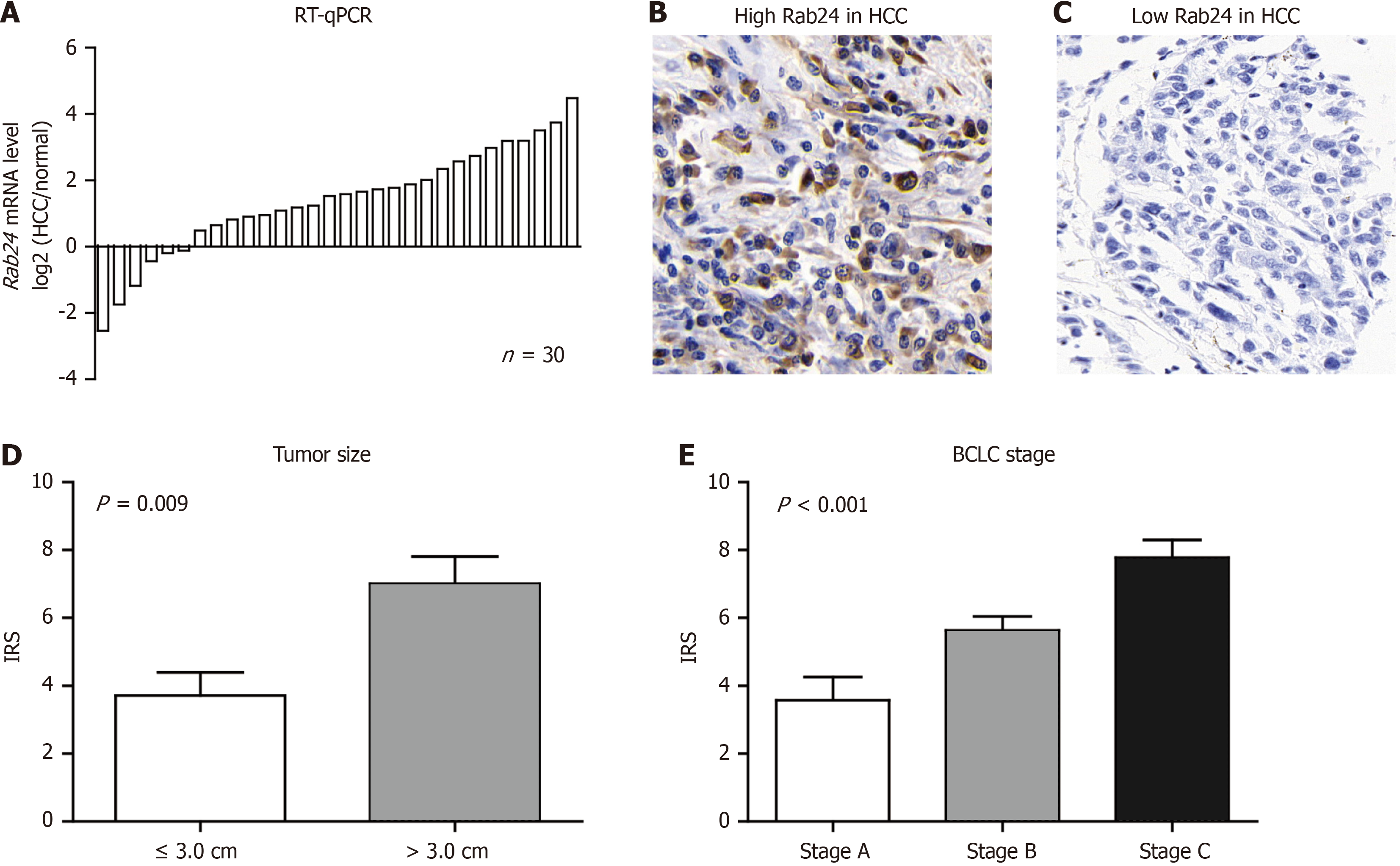
Figure 2 Expression of Rab24 in our enrolled hepatocellular carcinoma cohort.
A: Real-time PCR experiments were conducted to evaluate mRNA levels of Rab24 in clinical resected paired tissues (n = 30), exhibiting a significant upregulation in hepatocellular carcinoma (HCC) tissues compared to adjacent noncancerous liver tissues; B: Immunohistochemistry (IHC) staining results showed representative high expression level of Rab24 in HCC tissues [IHC immunoreactive score (IRS) = 12]. Magnification, 400 ×; C: Representative low Rab24 expression in HCC tissues (IRS = 0); D: Differences of Rab24 IRS between patients with small tumor size (≤ 3.0 cm, n = 51) or large tumor size (> 3.0 cm, n = 96). Data was shown as mean ± SD and analyze by Student’s t-test; E: Differences of Rab24 IRS among patients with different Barcelona Clinic Liver Cancer stages, including stage A (n = 57), stage B (n = 46), and stage C (n = 44). Data was shown as mean ± SD and analyze by One-way ANOVA test. BCLC: Barcelona Clinic Liver Cancer; RT-qPCR: Real-time PCR; HCC: Hepatocellular carcinoma.
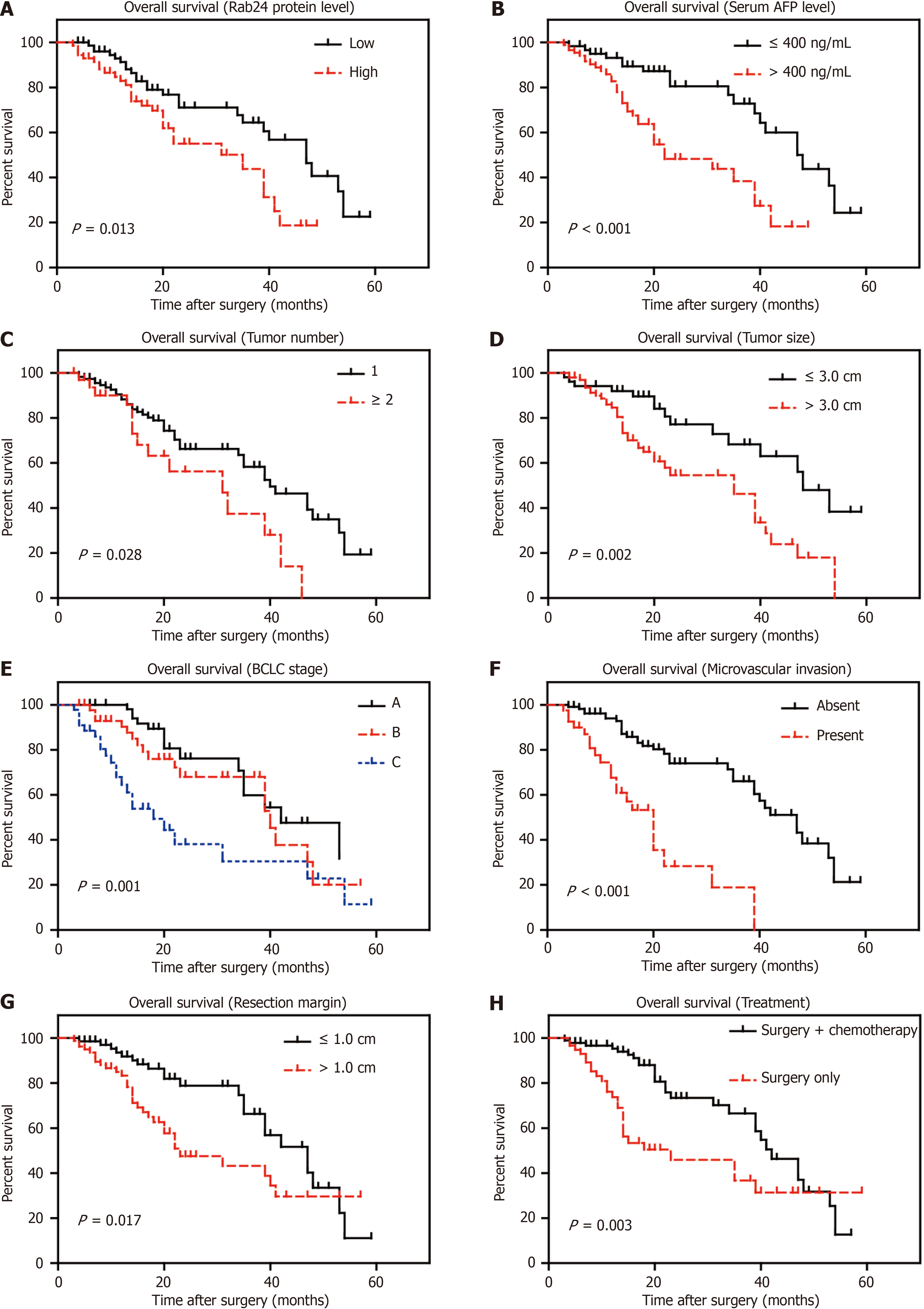
Figure 3 Survival analyses of hepatocellular carcinoma patients in our cohort.
Kaplan-Meier method was used to analyze the prognostic significance of all retrieved clinicopathological characteristics, which revealed that Rab24 expression, serum alpha-fetoprotein (AFP) level, tumor number, tumor size, Barcelona Clinic Liver Cancer (BCLC) stage, microvascular invasion, resection margin, and treatment therapy are all significant prognosis factors. A: Rab24 expression; B: Serum AFP level; C: Tumor number; D: Tumor size; E: BCLC stage; F: Microvascular invasion; G: Resection margin; H: Treatment therapy. Data was analyzed by log-rank test. BCLC: Barcelona Clinic Liver Cancer; AFP: Alpha-fetoprotein.
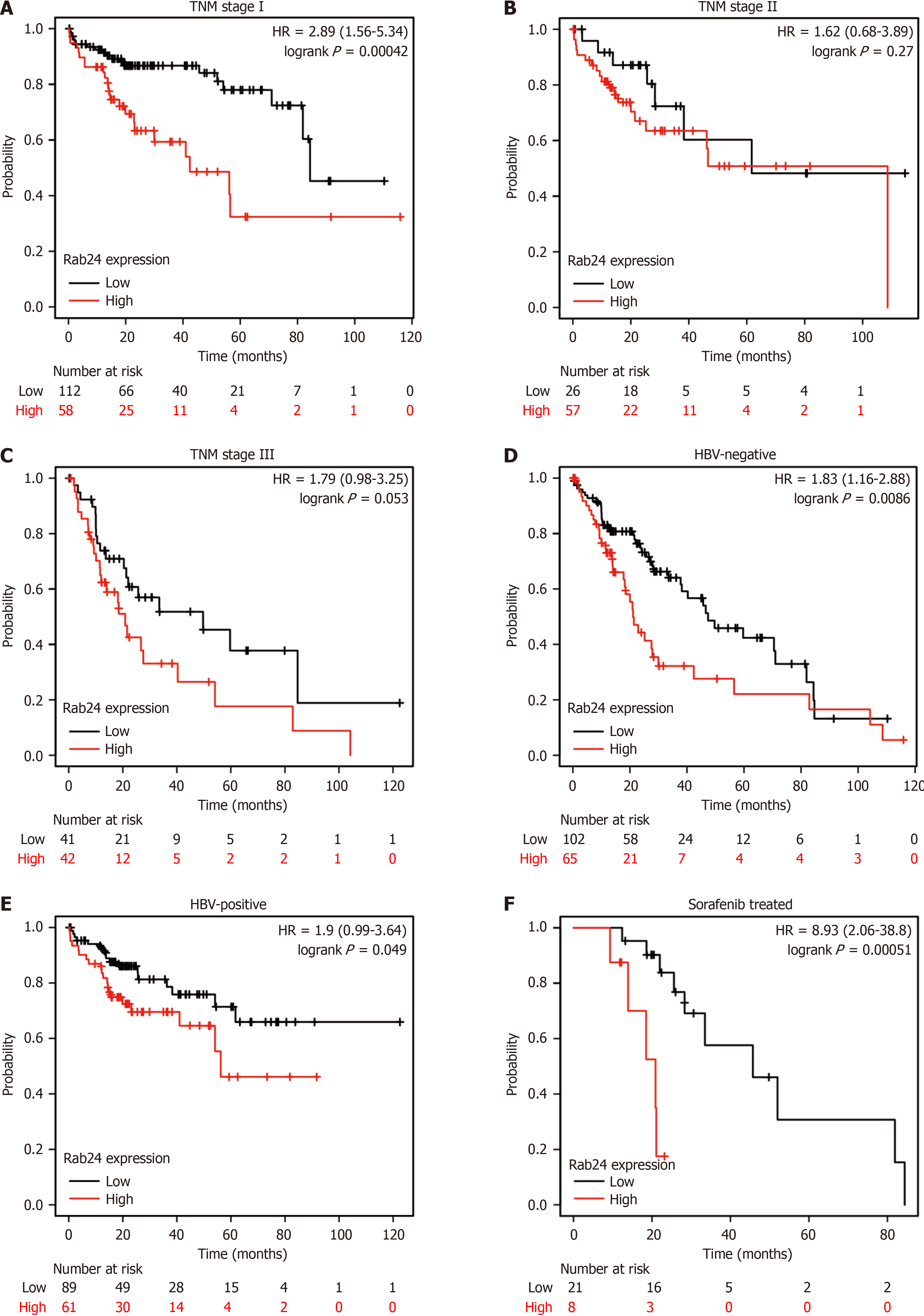
Figure 4 Stratification survival analyses of hepatocellular carcinoma patients.
A-C: Hepatocellular carcinoma (HCC) patients with tumor-node-metastasis stage I (A), stage II (B), or stage III (C) were independently analyzed to evaluate the prognostic effect of Rab24 level; D-F: Meanwhile, patients with negative-hepatitis B virus (HBV) infection history (D) or positive-HBV history (E) were also stratified, further revealing the unfavorable role of Rab24 in HCC patients (F). Patients who received chemotherapy using Sorafenib without surgical treatment were also analyzed to test the prognostic effect of Rab24. Data was analyzed by log-rank test using Kaplan-Meier method. HBV: Hepatitis B virus; TNM: Tumor-node-metastasis.
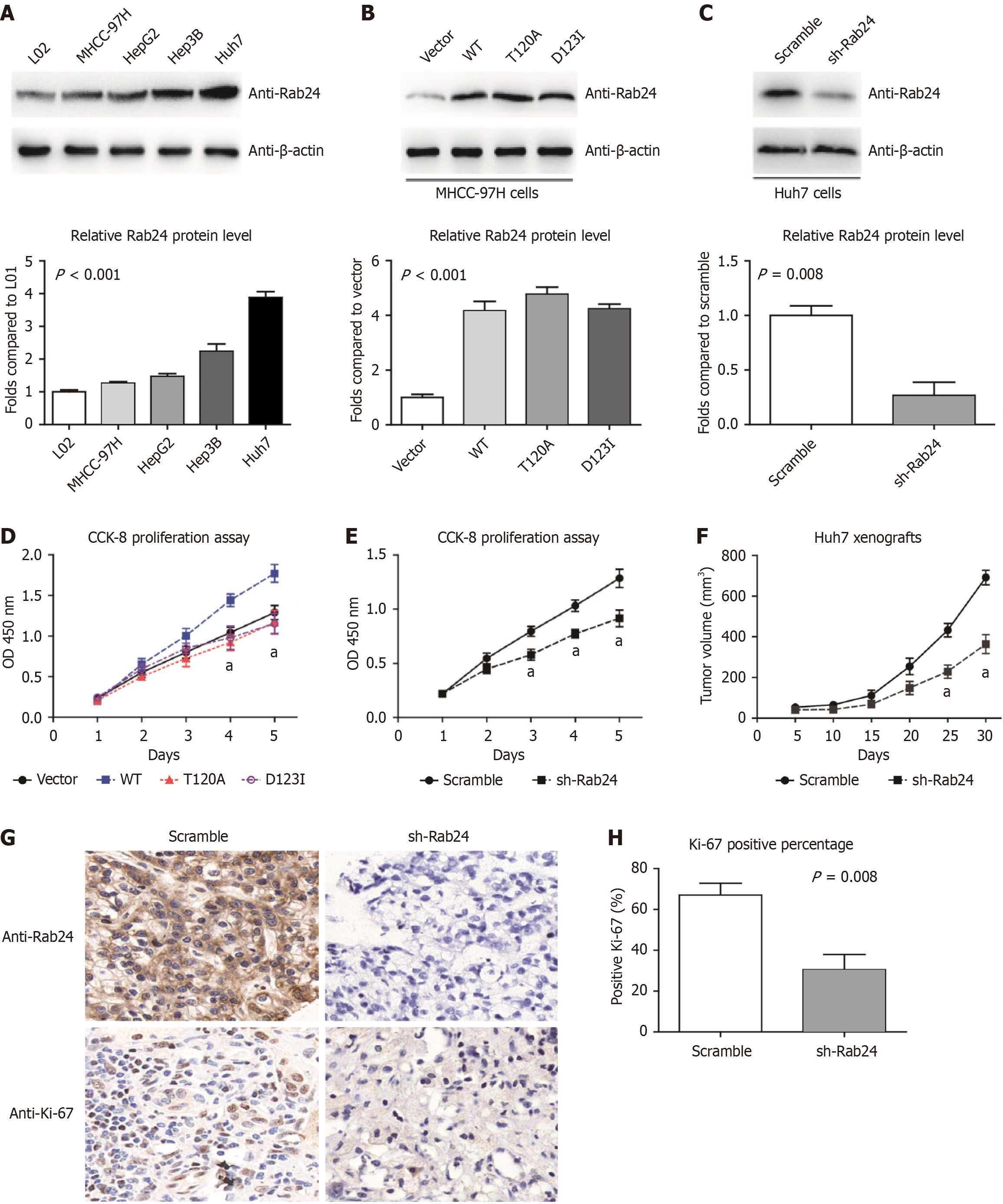
Figure 5 In vitro and in vivo evaluation of Rab24 function in hepatocellular carcinoma progression.
A: Western blotting data demonstrated higher Rab24 protein levels in human hepatocellular carcinoma (HCC) cell lines (Huh7, HepG2, Hep3B, and MHCC-97H) compared to noncancerous hepatocytes (L02); B: MHCC-97H cells were transfected with pcDNA3.1-vector, pcDNA3.1-Rab24-WT, pcDNA3.1-Rab24-T120A, or pcDNA3.1-Rab24-d123I plasmids for overexpression experiments; C: Knockdown assay was conducted by transfecting lentivirus shRNA targeting Rab24 in Huh7 cells, using scramble shRNA as control; D: Overexpressing Rab24-WT can significantly enhance the proliferation capacity of MHCC-97H cells, while its T120A or D123I mutants showed no significant difference compared to the control group transfected with pcDNA3.1-vector; E: Silencing Rab24 resulted in a significant inhibition on the proliferation process of Huh7 cells, as revealed by cell counting kit-8 assays; F: Huh7 cells transfected with Rab24-shRNA or scramble shRNA were subcutaneously injected into nude mice for xenograft assays. The tumor growth curve revealed that Rab24-knockdown significantly attenuated HCC growth in vivo; G: The isolated xenografts were tested by immunohistochemistry targeting Rab24 and Ki-67, respectively; H: Statistical analysis of Ki-67 immunoreactivity showed a significant lower Ki-67 Level in xenografts with Rab24-knockdown. Data was shown as mean ± SD and analyze by Student’s t-test. aP < 0.05; CCK-8: Cell counting kit-8; WT: Wild type.
- Citation: Ding H, Ding ZG, Liu S, Mao XN, Lu XS. Ras-related protein Rab24 plays a predictive role in hepatocellular carcinoma and enhanced tumor proliferation. World J Gastroenterol 2025; 31(8): 101585
- URL: https://www.wjgnet.com/1007-9327/full/v31/i8/101585.htm
- DOI: https://dx.doi.org/10.3748/wjg.v31.i8.101585


