Copyright
©The Author(s) 2025.
World J Gastroenterol. Feb 14, 2025; 31(6): 96782
Published online Feb 14, 2025. doi: 10.3748/wjg.v31.i6.96782
Published online Feb 14, 2025. doi: 10.3748/wjg.v31.i6.96782
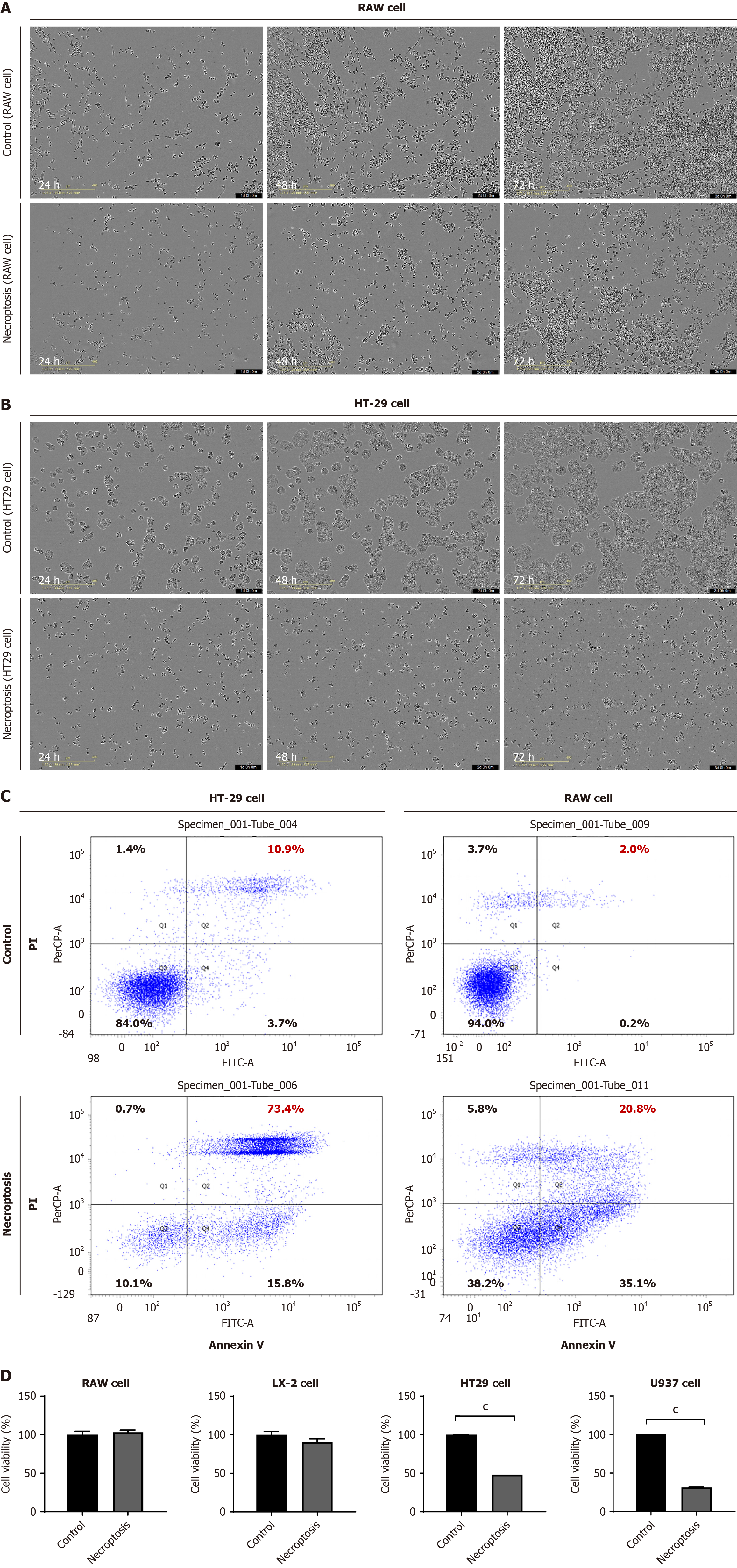
Figure 1 Cell death after necroptosis stimuli differed according to cell type.
A: After inducing necroptosis in RAW 264.7 cells, cell viability was determined using Incucyte assay; B: After inducing necroptosis in HT29 cells, cell viability was detected using Incucyte assay; C: Flow cytometry shows the ratio of necrosis induced in RAW 264.7 and HT9 cells after necroptosis induction; D: Cell viability was detected after necroptosis. Results are shown as mean. cP < 0.001. PI: Propidium iodide.
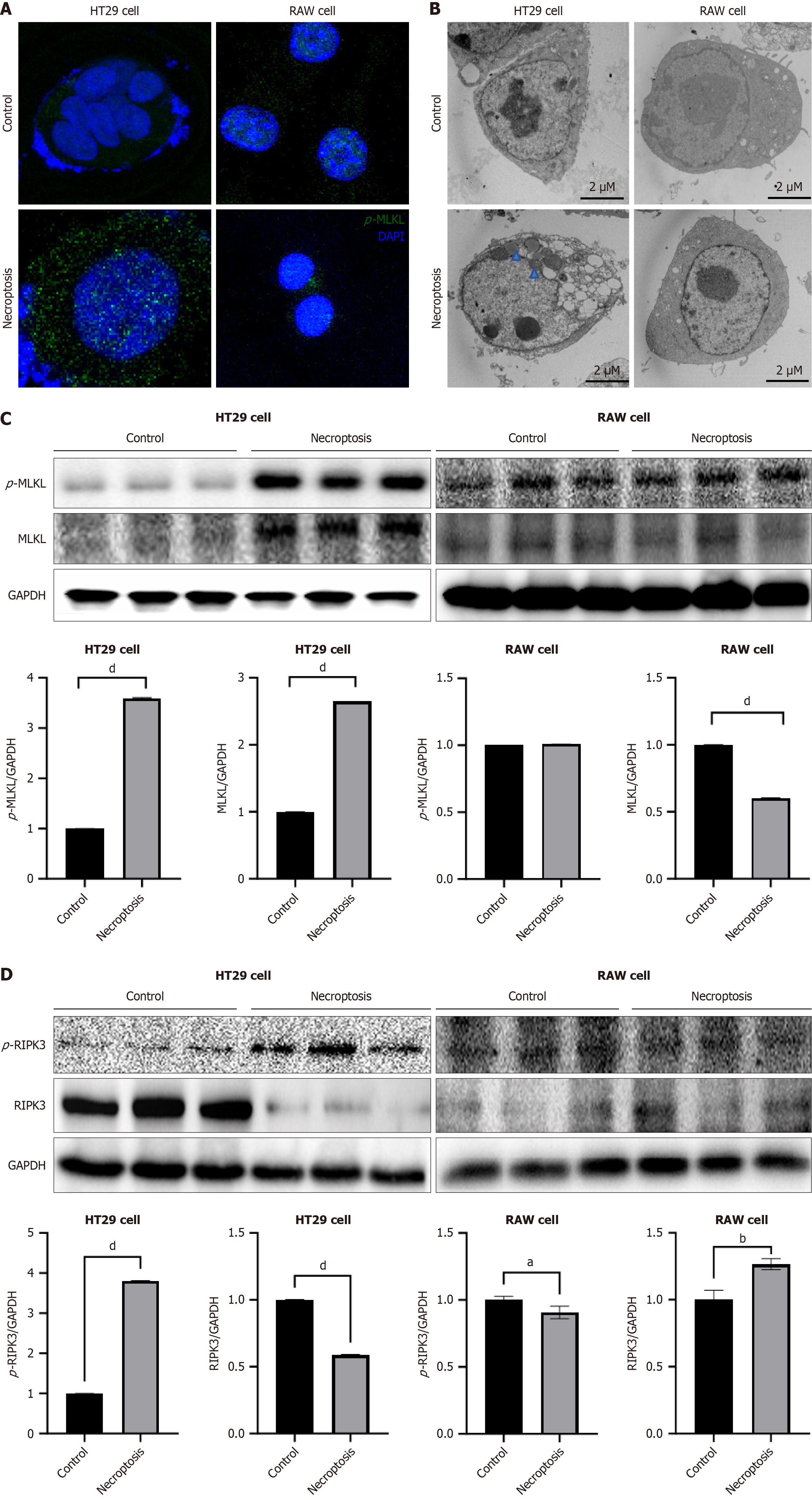
Figure 2 RAW cells did not phosphorylate mixed lineage kinase domain-like protein and avoided cell death after necroptosis stimuli.
A: Fixed and permeabilized HT29 and RAW 264.7 cells stained with 4’,6-diamidino-2-phenylindole were imaged by confocal microscopy before necroptosis induced [antibody: Phosphorylated-mixed lineage kinase domain-like protein (p-MLKL)]; B: Representative transmission electron microscopy images of HT29 and RAW 264.7 cells undergoing necroptosis for 24 hours. The blue arrow represents mitochondria, whereas the black arrow represents the plasma membrane (Scale bars: 2 μm); C: Western blot analysis of p-MLKL and MLKL protein expression in HT29 and RAW 264.7 cells before necroptosis; D: Western blot analysis of phosphorylated-receptor-interacting serine/threonine kinase 3 (RIPK3) and RIPK3 protein expression in HT29 and RAW 264.7 cells after necroptosis. aP < 0.05. bP < 0.01. dP < 0.0001. p-MLKL: Phosphorylated-mixed lineage kinase domain-like protein; DAPI: 4’,6-Diamidino-2-phenylindole dihydrochloride; MLKL: Mixed lineage kinase domain-like protein; RIPK3: Receptor-interacting serine/threonine kinase 3; p-RIPK3: Phosphorylated-receptor-interacting serine/threonine kinase 3; GAPDH: Glyceraldehyde 3-phosphate dehydrogenase.
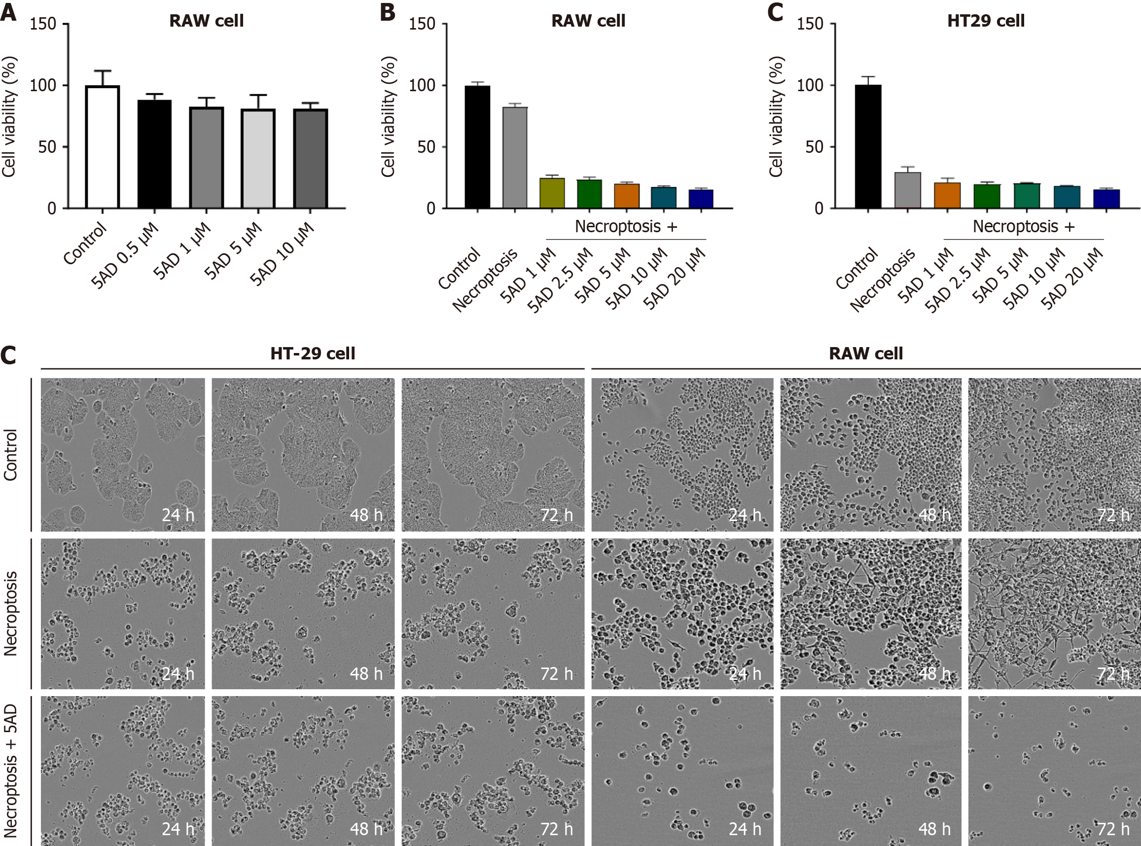
Figure 3 Necroptosis-induced cell death occurs after demethylation in RAW 264.
7 cell. A: RAW 264.7 cells treated with 5-aza-2’-deoxycytidine (5AD) to determine cell cytotoxicity; B: In HT29 and RAW 264.7 cells, cell viability was tested via demethylation with 5AD for 72 hours and induction of necroptosis for 24 hours; C: After inducing necroptosis in HT29 and RAW 264.7 cells, cell viability was determined using Incucyte assay. 5AD: 5-aza-2’-deoxycytidine.
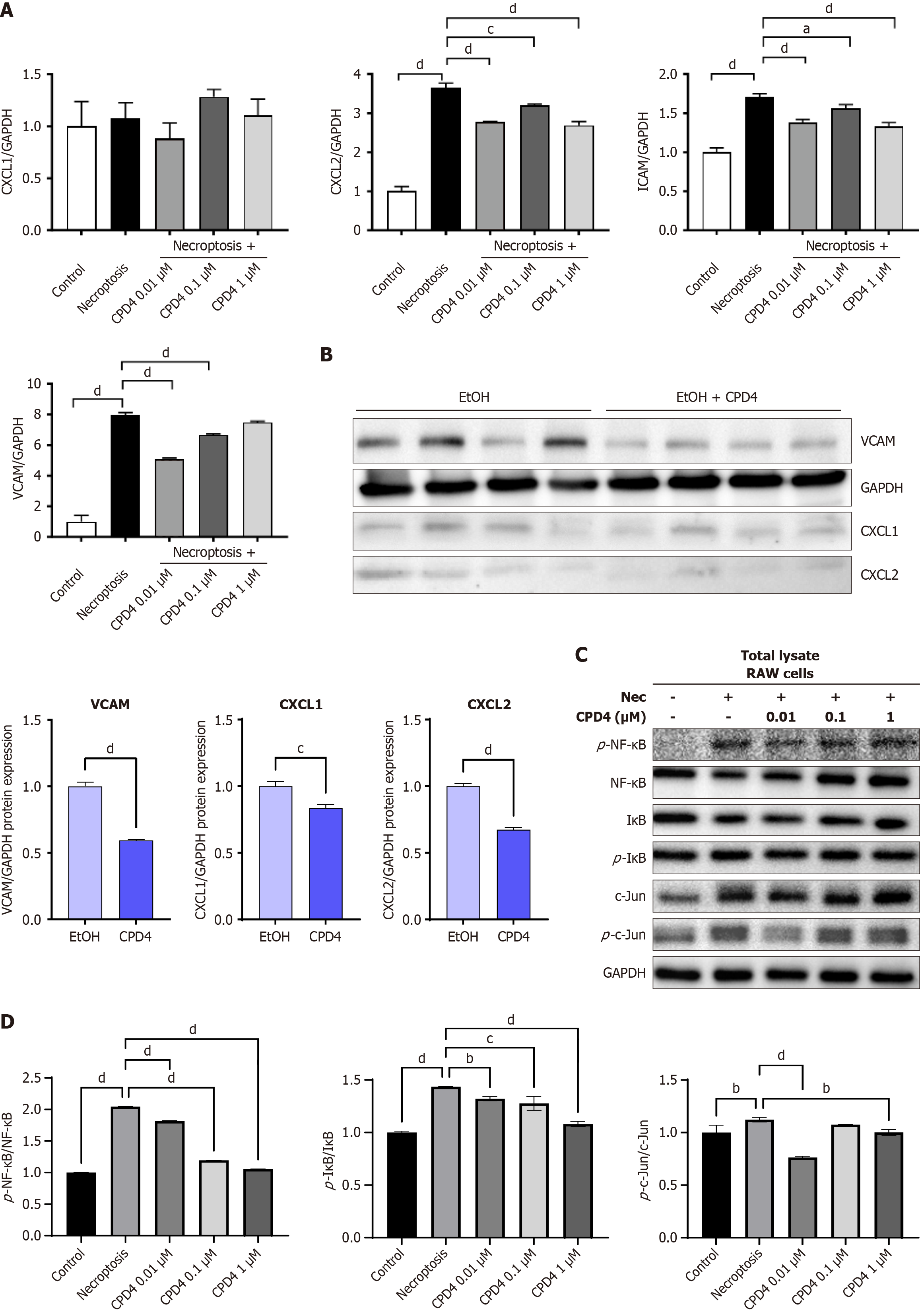
Figure 4 Mixed lineage kinase domain-like protein adenosine triphosphate-binding inhibitor cannot evade cell death after necroptosis stimuli but inhibition of inflammation.
A: U937, HT29 and FADD cells treated with Mixed lineage kinase domain-like protein (MLKL) adenosine triphosphate (ATP)-binding inhibitor (Compound-4) to determine cell viability; B: After induction of necroptosis in RAW 264.7 cells, MLKL ATP-binding inhibitor was administered to measure CXCL1, CXCL2, ICAM, and VCAM mRNA expression levels via quantitative real-time polymerase chain reaction; C and D: Western blot analysis of phosphorylated-nuclear factor kappa-B, nuclear factor kappa-B, phospho-c-Jun, c-Jun, I-kappa-B-alpha, phospho-I-kappa-B-alpha, c-Jun N-terminal kinases and phospho-c-Jun N-terminal kinases protein expression in RAW 264.7 cells after compound-4 treatment. aP < 0.05. bP < 0.01. cP < 0.001. dP < 0.0001. GAPDH: Glyceraldehyde 3-phosphate dehydrogenase; CPD4: Compound-4; EtOH: Ethyl alcohol.
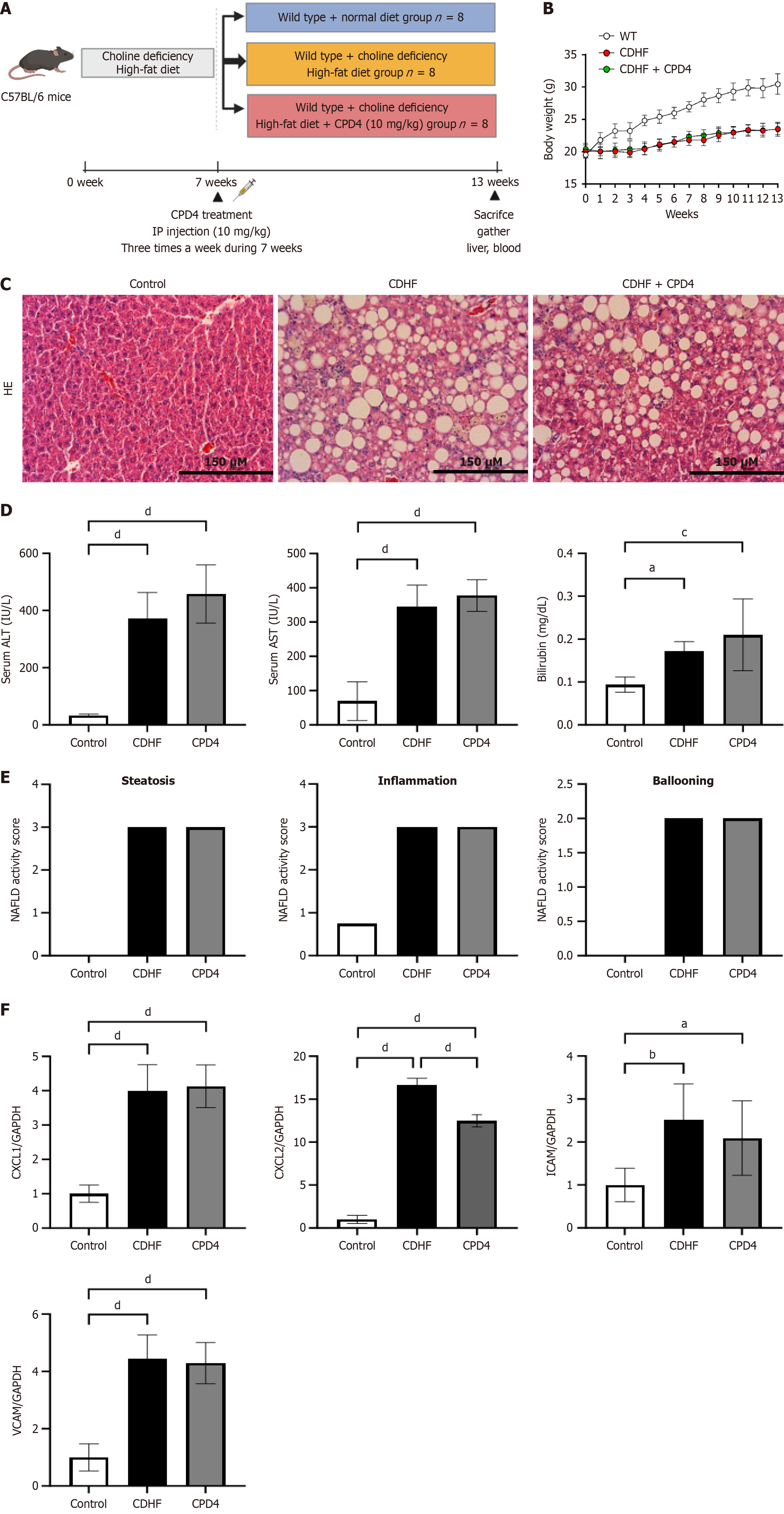
Figure 5 Mixed lineage kinase domain-like protein adenosine triphosphate pocket-binding inhibitor could not attenuate steatosis and inflammation in non-alcoholic fatty liver disease animal model.
A: Overview of animal experiment. Mice were fed choline-deficient high-fat diet for 13 weeks. After six weeks, mixed lineage kinase domain-like protein (MLKL) adenosine triphosphate (ATP)-binding inhibitor (Compound-4) (10 mg/kg) was injected intraperitoneally three times a week for a total of seven weeks; B: C57BL/6 mouse body weight through the 13 weeks of experiment. No significant difference was evident between choline-deficient high-fat (CDHF) and CDHF + MLKL ATP-binding inhibitor (Compound-4); n = 8; C: Liver tissue sections were stained with hematoxylin-eosin staining, 200 ×; D: Serum alanine transaminase, aspartate transaminase, and total bilirubin were measured (n = 8/group); E: Steatosis, inflammation, and hepatocyte ballooning among control, CDHF, and MLKL ATP-binding inhibitor (Compound-4) groups; F: CXCL1, CXCL2, ICAM, and VCAM mRNA expression levels were measured via quantitative real-time polymerase chain reaction in liver tissue. P calculated compared to control, choline-deficient high-fat, mixed lineage kinase domain-like protein adenosine triphosphate binding inhibitor (Compound-4). aP < 0.05. bP < 0.01. cP < 0.001. dP < 0.0001. HE: Hematoxylin-eosin staining; CDHF: Choline-deficient high-fat; GAPDH: Glyceraldehyde 3-phosphate dehydrogenase; CPD4: Compound-4; ALT: Alanine transaminase; AST: Aspartate transaminase; NAFLD: Non-alcoholic fatty liver disease.
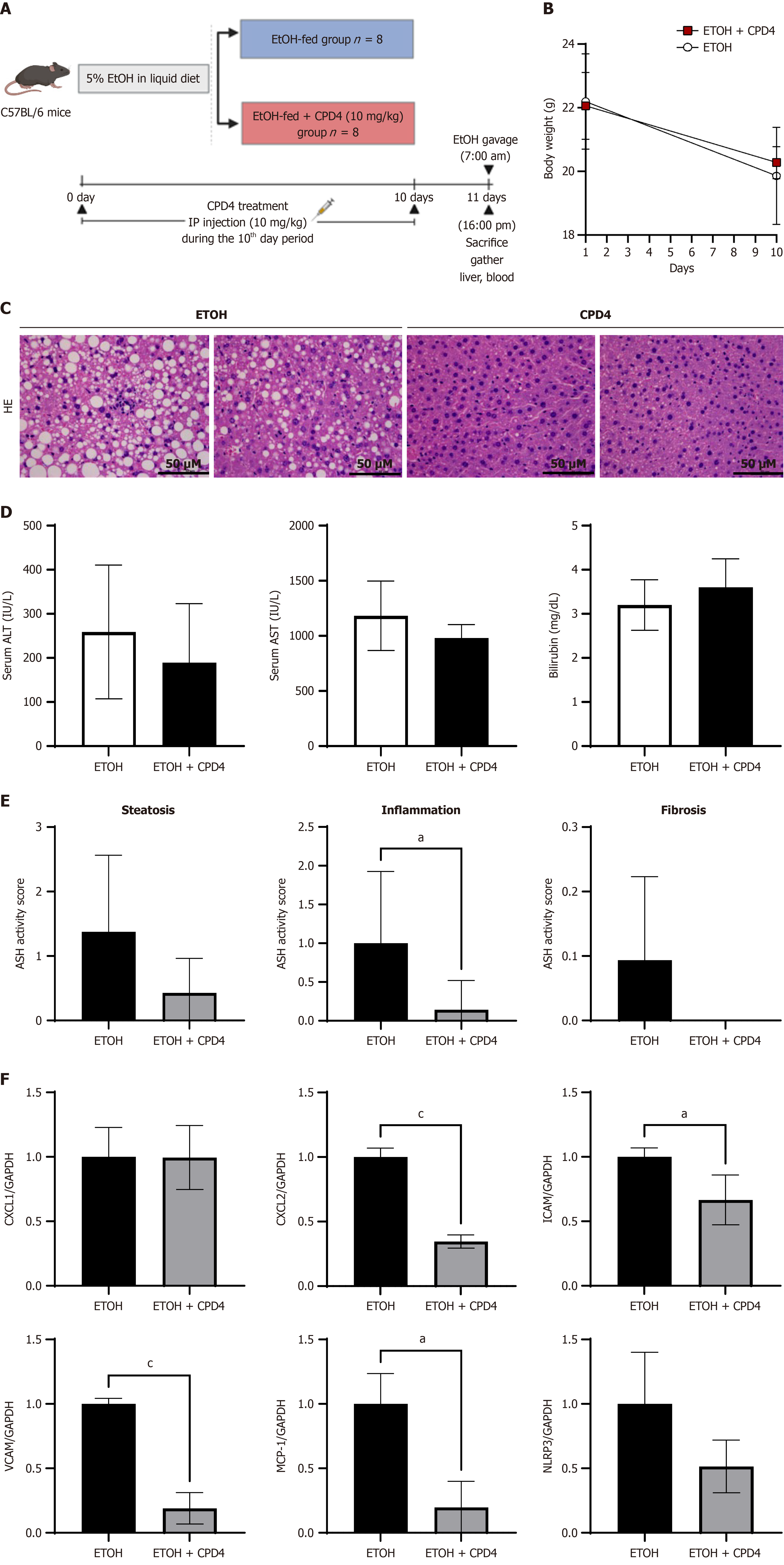
Figure 6 Mixed lineage kinase domain-like protein adenosine triphosphate-binding inhibitor attenuated steatosis and inflammation in alcoholic animal model.
A: Overview of animal experiments. Mice were fed an ethyl alcohol (EtOH) diet for 10 days, and daily intraperitoneal injection of mixed lineage kinase domain-like protein (MLKL) adenosine triphosphate (ATP)-binding inhibitor (Compound-4) (10 mg/kg) was administered for 10 days. On the morning of 11th day, EtOH was administered orally (5 g/kg), and euthanasia was performed nine hours later. Liver tissue and serum were collected; B: C57BL/6 mouse body weight through 10 days of experiment. The mice fed EtOH diet showed a trend of weight loss; n = 8; C: Liver tissue sections were stained with hematoxylin-eosin staining, 200 ×; D: Serum alanine transaminase, aspartate transaminase, and total bilirubin were measured (n = 8/group); E: Steatosis, inflammation, and fibrosis among EtOH and MLKL ATP-binding inhibitor (Compound-4) groups; F: CXCL1, CXCL2, ICAM, VCAM, MCP-1, and NLRP3 mRNA expression levels were measured via quantitative real-time polymerase chain reaction in liver tissue. P calculated compared to ethyl alcohol, mixed lineage kinase domain-like protein adenosine triphosphate-binding inhibitor (Compound-4). aP < 0.05. cP < 0.001. EtOH: Ethyl alcohol; HE: Hematoxylin-eosin staining; GAPDH: Glyceraldehyde 3-phosphate dehydrogenase; CPD4: Compound-4; ALT: Alanine transaminase; AST: Aspartate transaminase; ASH: Alcoholic steatohepatitis.
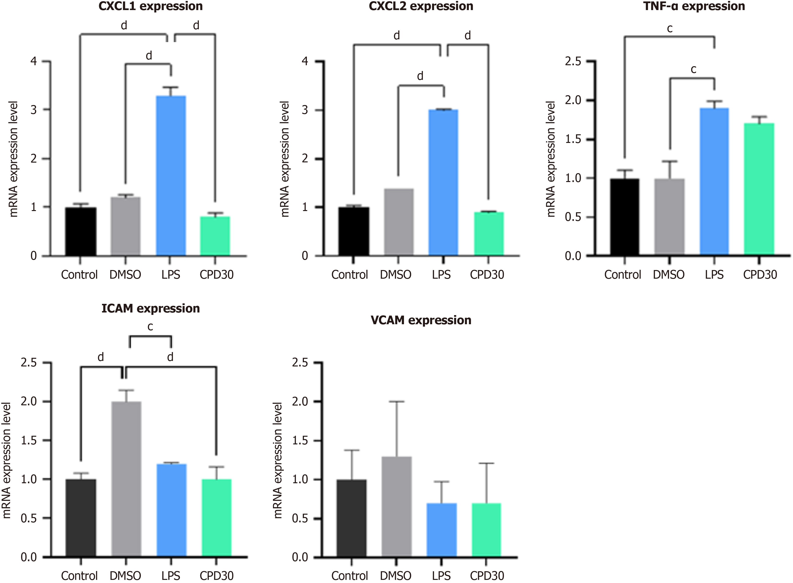
Figure 7 Effects of mixed lineage kinase domain-like protein adenosine triphosphate pocket inhibitor on alcoholic liver disease model after lipopolysaccharide stimuli in liver organoid system.
Liver organoids were treated with lipopolysaccharide to create an alcohol-induced liver disease model. After mixed lineage kinase domain-like protein adenosine triphosphate pocket-binding inhibitor treatment after necroptotic stimuli, the expression of tumor necrosis factor-α, CXCL1/CXCL2, ICAM, and VCAM decreased in the organoid system. cP < 0.001. dP < 0.0001. TNF: Tumor necrosis factor; DMSO: Dimethyl sulfoxide; LPS: Lipopolysaccharide; CPD: Compound.
- Citation: Xuan Yuan HN, Kim HS, Park GR, Ryu JE, Kim JE, Kang IY, Kim HY, Lee SM, Oh JH, Yoon EL, Jun DW. Adenosine triphosphate-binding pocket inhibitor for mixed lineage kinase domain-like protein attenuated alcoholic liver disease via necroptosis-independent pathway. World J Gastroenterol 2025; 31(6): 96782
- URL: https://www.wjgnet.com/1007-9327/full/v31/i6/96782.htm
- DOI: https://dx.doi.org/10.3748/wjg.v31.i6.96782


