Copyright
©The Author(s) 2025.
World J Gastroenterol. Feb 14, 2025; 31(6): 102070
Published online Feb 14, 2025. doi: 10.3748/wjg.v31.i6.102070
Published online Feb 14, 2025. doi: 10.3748/wjg.v31.i6.102070
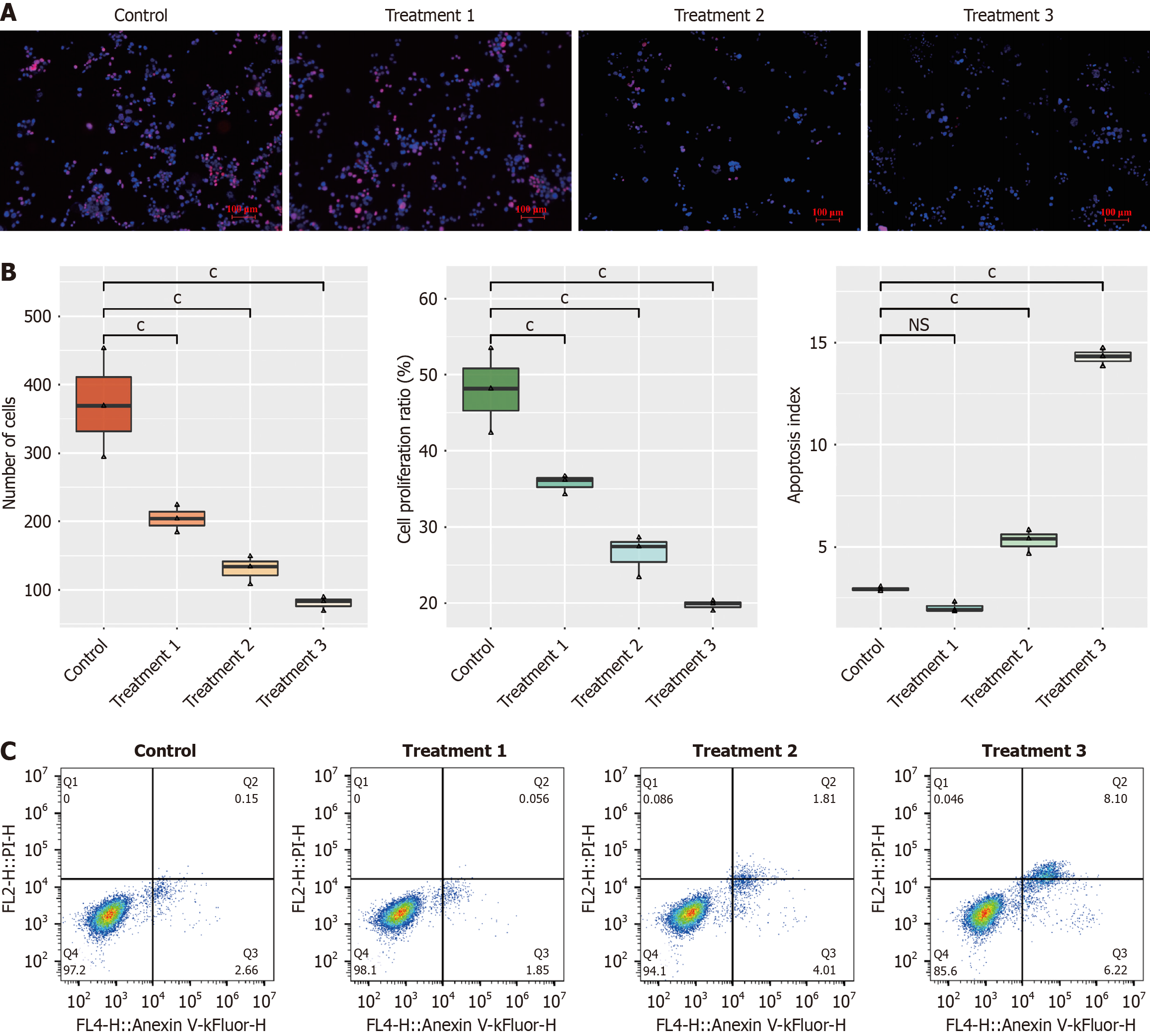
Figure 1 Effect of different concentrations of keratin 1 antibody on Caco-2 cell proliferation and apoptosis.
A and B: Keratin 1 (KRT1) antibody inhibited cellular proliferation; C: KRT1 antibody induced apoptosis. cP < 0.001. P value vs control group. Control refers to untreated Caco-2 cells; Treatment 1: Keratin 1 (KRT1) antibody at 1 ng/mL; Treatment 2: KRT1 antibody at 5 ng/mL; Treatment 3: KRT1 antibody at 10 ng/mL. Same for subsequent figures.
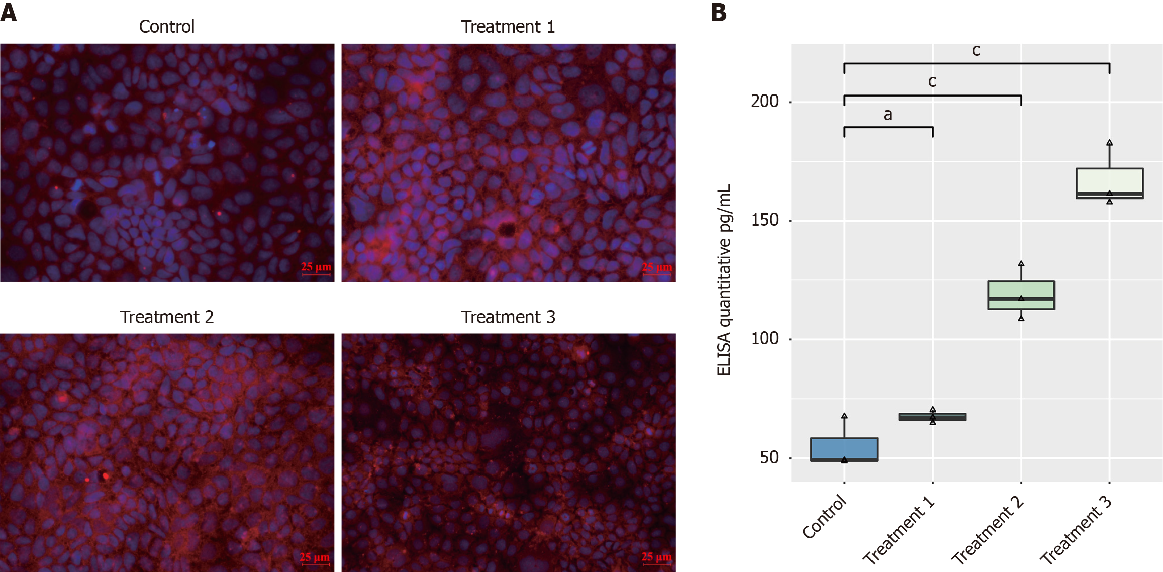
Figure 2 Effect of different concentrations of keratin 1 antibody on the expression levels of high molecular weight kininogen and bradykinin in Caco-2 cells.
A: Immunofluorescence analysis shows a reduction in high molecular weight kininogen expression in Caco-2 cells treated with different concentrations of keratin 1 (KRT1) antibody; B: Enzyme-linked immunosorbent assay results demonstrate an increase in bradykinin expression in Caco-2 cells treated with various concentrations of KRT1 antibody. aP < 0.05. cP < 0.001. P value vs control group. Treatment 1: Keratin 1 (KRT1) antibody at 1 ng/mL; Treatment 2: KRT1 antibody at 5 ng/mL; Treatment 3: KRT1 antibody at 10 ng/mL. ELISA: Enzyme-linked immunosorbent assay.
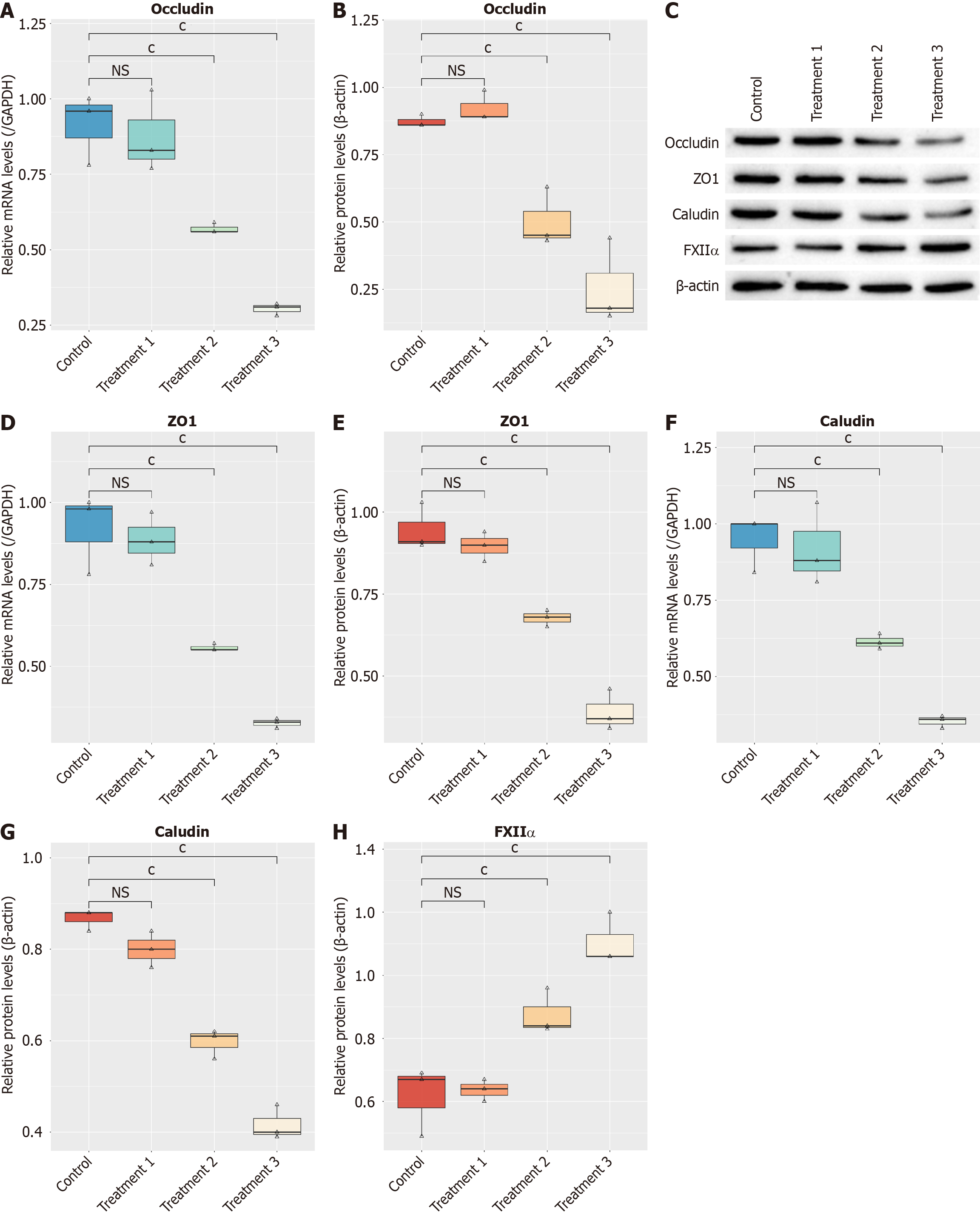
Figure 3 Effects of different concentrations of the keratin 1 antibody on intestinal mechanical barrier marker genes in Caco-2 cells.
A: Real-time polymerase chain reaction (RT-qPCR) results showed a reduction in the expression levels of occludin treated with varying concentrations of the keratin 1 (KRT1) antibody; B: Western blot quantification revealed decreased expression of occludin in Caco-2 cells treated with the KRT1 antibody; C: Western blot analysis of occludin, zonula occludens-1, claudin, and coagulation factor XII in Caco-2 cells under different treatment conditions; D: RT-qPCR results showed a reduction in the expression levels of zonula occludens-1 treated with varying concentrations of the KRT1 antibody; E: Western blot quantification revealed decreased expression of zonula occludens-1 in Caco-2 cells treated with the KRT1 antibody; F: RT-qPCR results showed a reduction in the expression levels of claudin in Caco-2 cells treated with varying concentrations of the KRT1 antibody; G: Western blot quantification revealed decreased expression of claudin in Caco-2 cells treated with the KRT1 antibody; H: Western blot quantification increased expression of coagulation factor XII, in Caco-2 cells treated with the KRT1 antibody. cP < 0.001. P value vs control group. Treatment 1: Keratin 1 (KRT1) antibody at 1 ng/mL; Treatment 2: KRT1 antibody at 5 ng/mL; Treatment 3: KRT1 antibody at 10 ng/mL. ZO-1: Zonula occludens-1; FXIIα: Coagulation factor XII; GAPDH: Glyceraldehyde-3-phosphate dehydrogenase.
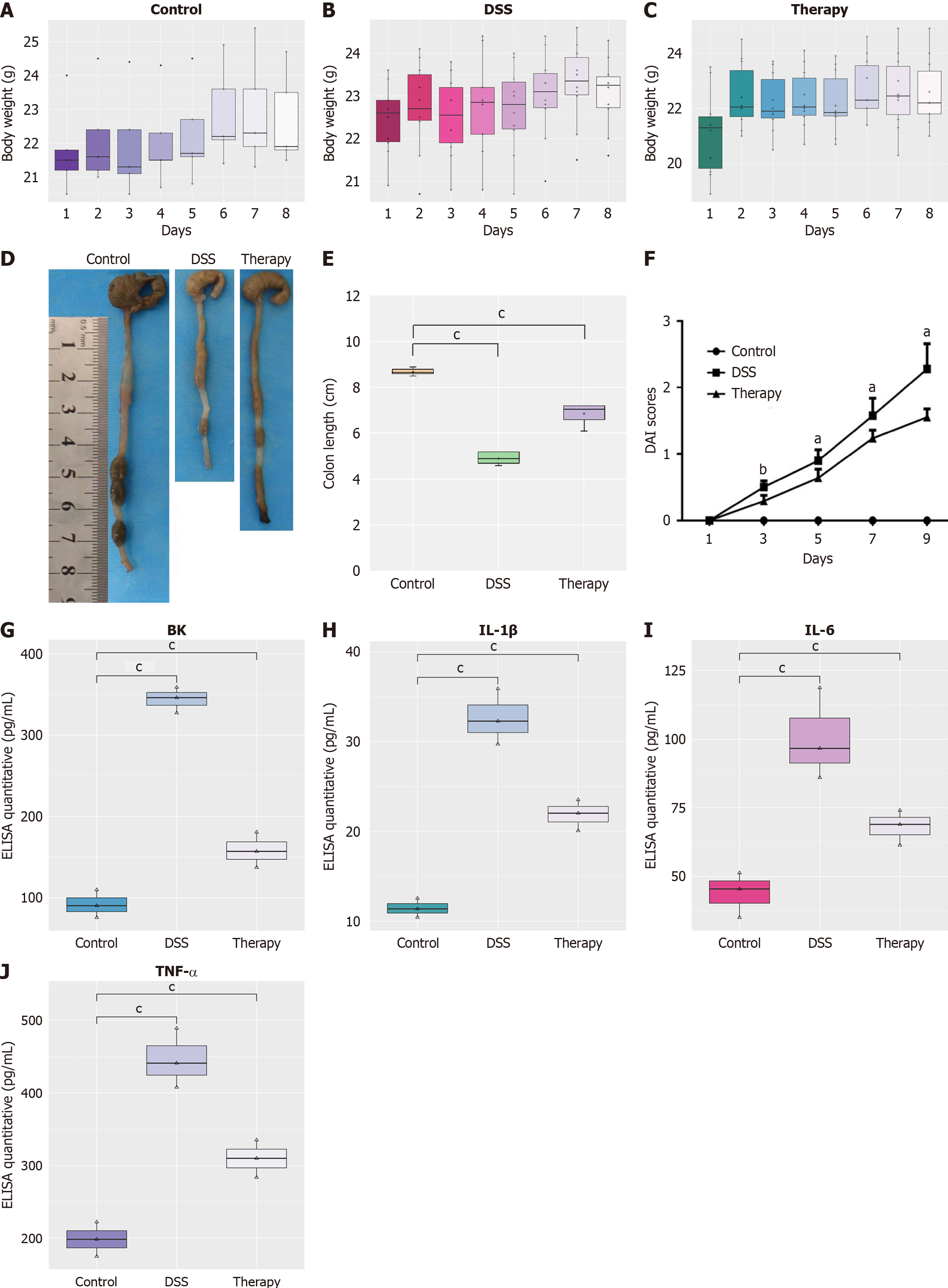
Figure 4 Anti-inflammatory effects of keratin 1 protein in mice with dextran sulfate sodium-induced colitis.
A-C: Relative body weight changes; D: Representative images of colons; E: Colon length measurements for each group; F: Disease activity index score; G: Enzyme-linked immunosorbent assay (ELISA) quantitative of bradykinin; H: ELISA quantitative of interleukin-1β; I: ELISA quantitative of interleukin-6; J: ELISA quantitative of tumor necrosis factor-α. aP < 0.05. bP < 0.01. cP < 0.001. P value vs control group. DSS: Dextran sulfate sodium; DAI: Disease activity index; ELISA: Enzyme-linked immunosorbent assay; BK: Bradykinin; IL: Interleukin; TNF: Tumor necrosis factor.
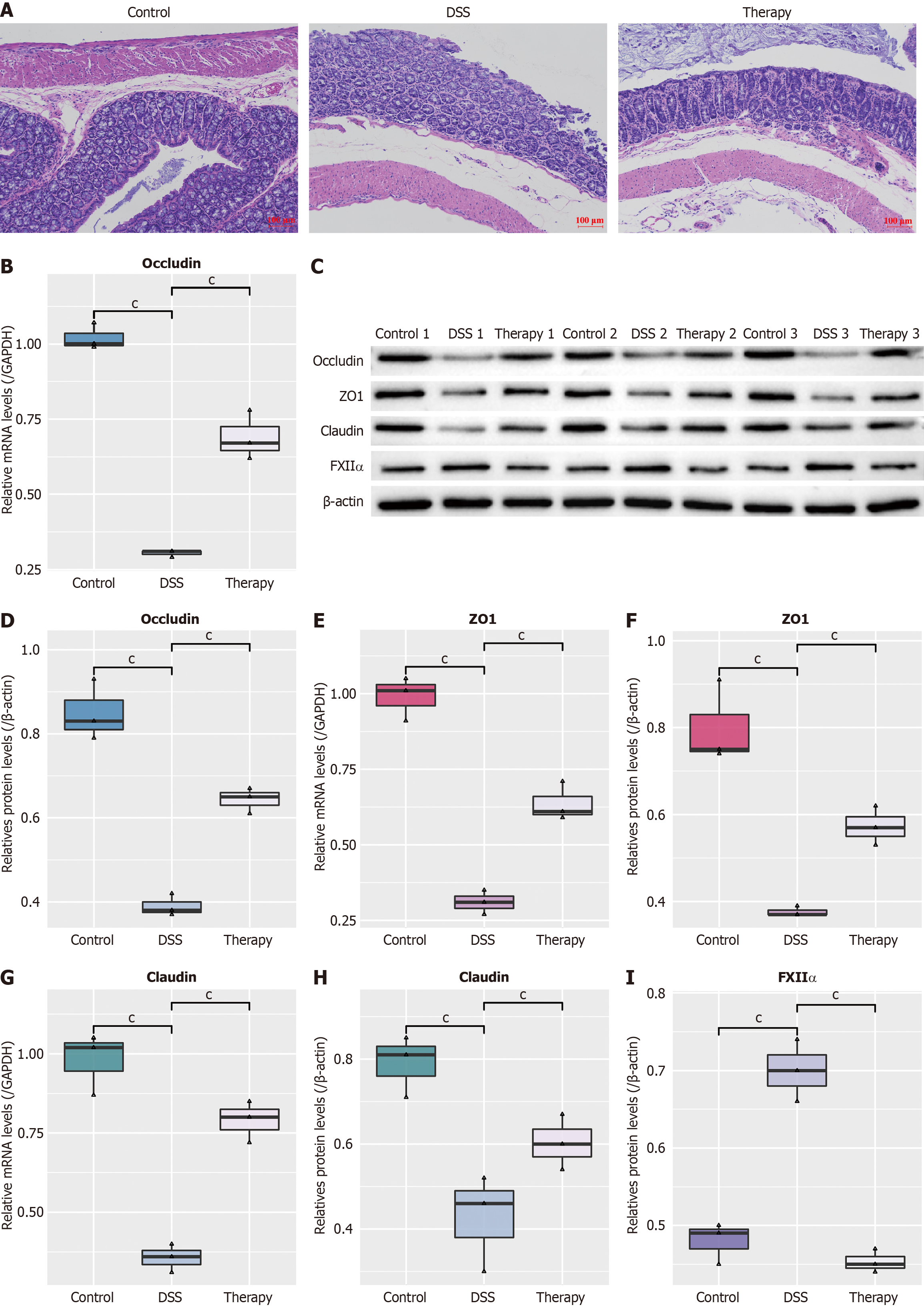
Figure 5 Intestinal changes and alterations in intestinal mechanical barrier marker gene expression in different treatment groups.
A: Hematoxylin-eosin staining of intestinal cross-sections from different treatment groups (100 ×); B: Real-time polymerase chain reaction (RT-qPCR) detection results of occludin expression in different groups; C: Western blot results of occludin, zonula occludens-1, claudin, and coagulation factor XII in different groups; D: Grayscale quantification of Western blot data for occludin in different groups; E: RT-qPCR detection results of zonula occludens-1 in different groups; F: Grayscale quantification of Western blot data for zonula occludens-1 in different groups; G: RT-qPCR detection results of claudin expression in different groups; H: Grayscale quantification of Western blot data for claudin in different groups; I: Grayscale quantification of Western blot data for coagulation factor XII in different groups. DSS: Dextran sulfate sodium; ZO-1: Zonula occludens-1; FXIIα: Coagulation factor XII; GAPDH: Glyceraldehyde-3-phosphate dehydrogenase.
- Citation: Dong XQ, Zhang YH, Luo J, Li MJ, Ma LQ, Qi YT, Miao YL. Keratin 1 modulates intestinal barrier and immune response via kallikrein kinin system in ulcerative colitis. World J Gastroenterol 2025; 31(6): 102070
- URL: https://www.wjgnet.com/1007-9327/full/v31/i6/102070.htm
- DOI: https://dx.doi.org/10.3748/wjg.v31.i6.102070


