Copyright
©The Author(s) 2025.
World J Gastroenterol. Jan 28, 2025; 31(4): 98752
Published online Jan 28, 2025. doi: 10.3748/wjg.v31.i4.98752
Published online Jan 28, 2025. doi: 10.3748/wjg.v31.i4.98752
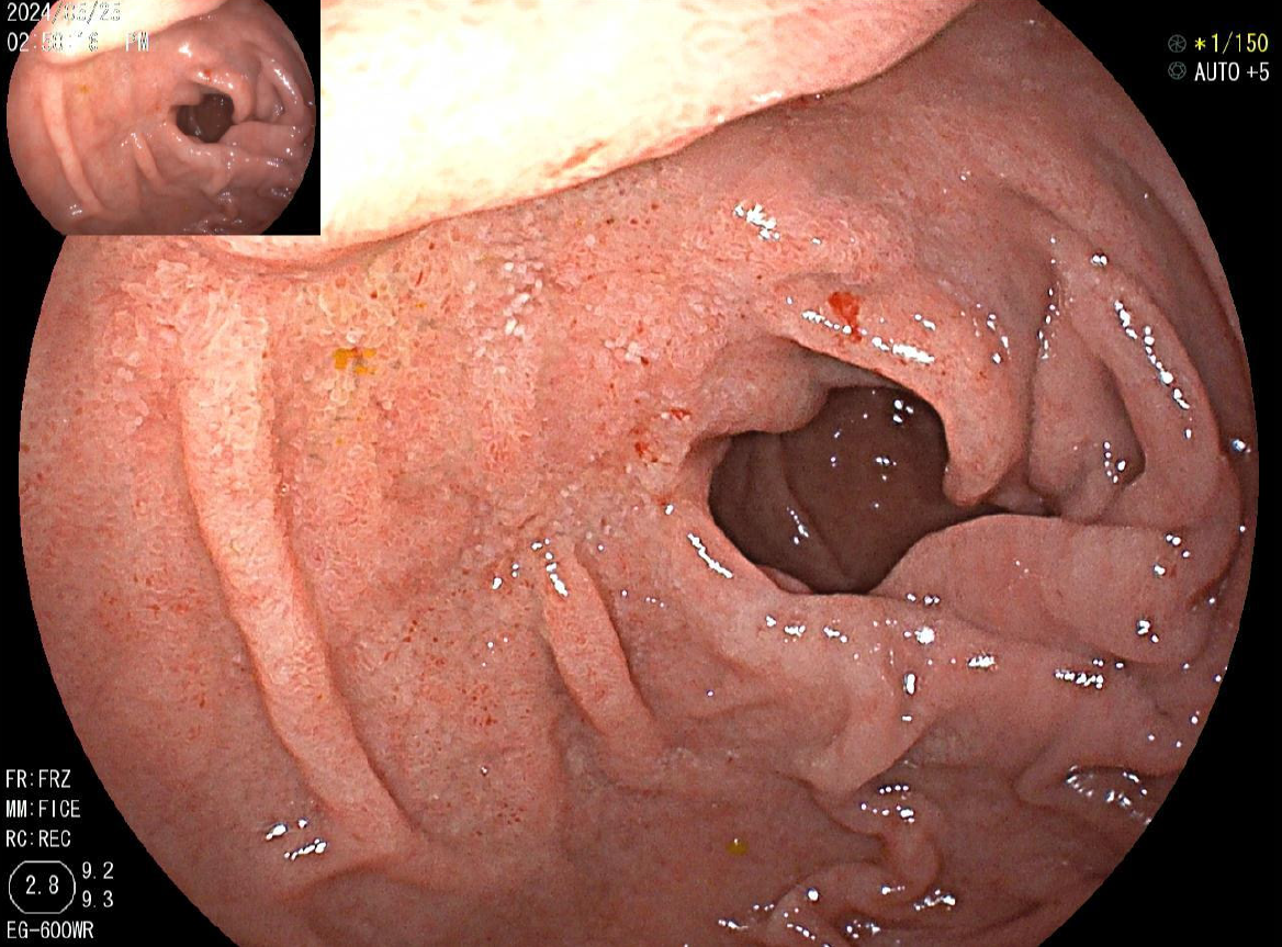
Figure 1 Endoscopy revealed diffuse erosions in the duodenum.
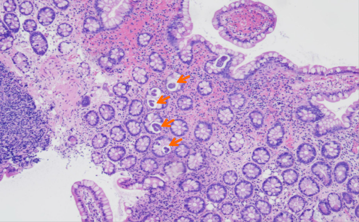
Figure 2 Strong affinity for basophilic staining (arrow) in the glandular cavity indicating the presence of eggs and immature forms of Strongyloides stercoralis.
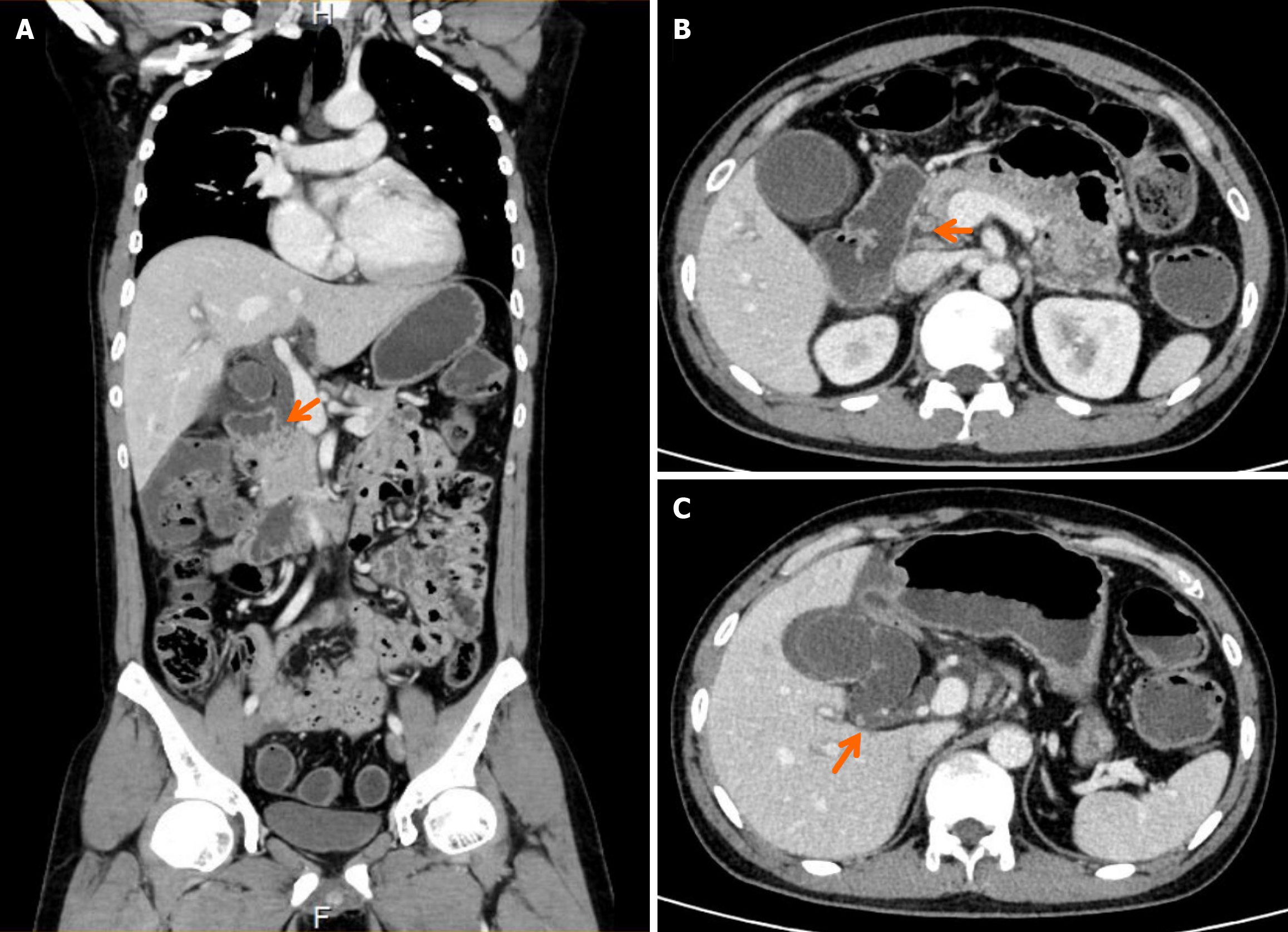
Figure 3 Computed tomography scan showing a gallstone obstructing the lower portion of the common bile duct and a gallbladder stone.
A and B: Gallstone (arrow) in the common bile duct; C: Gallbladder stone (arrow).
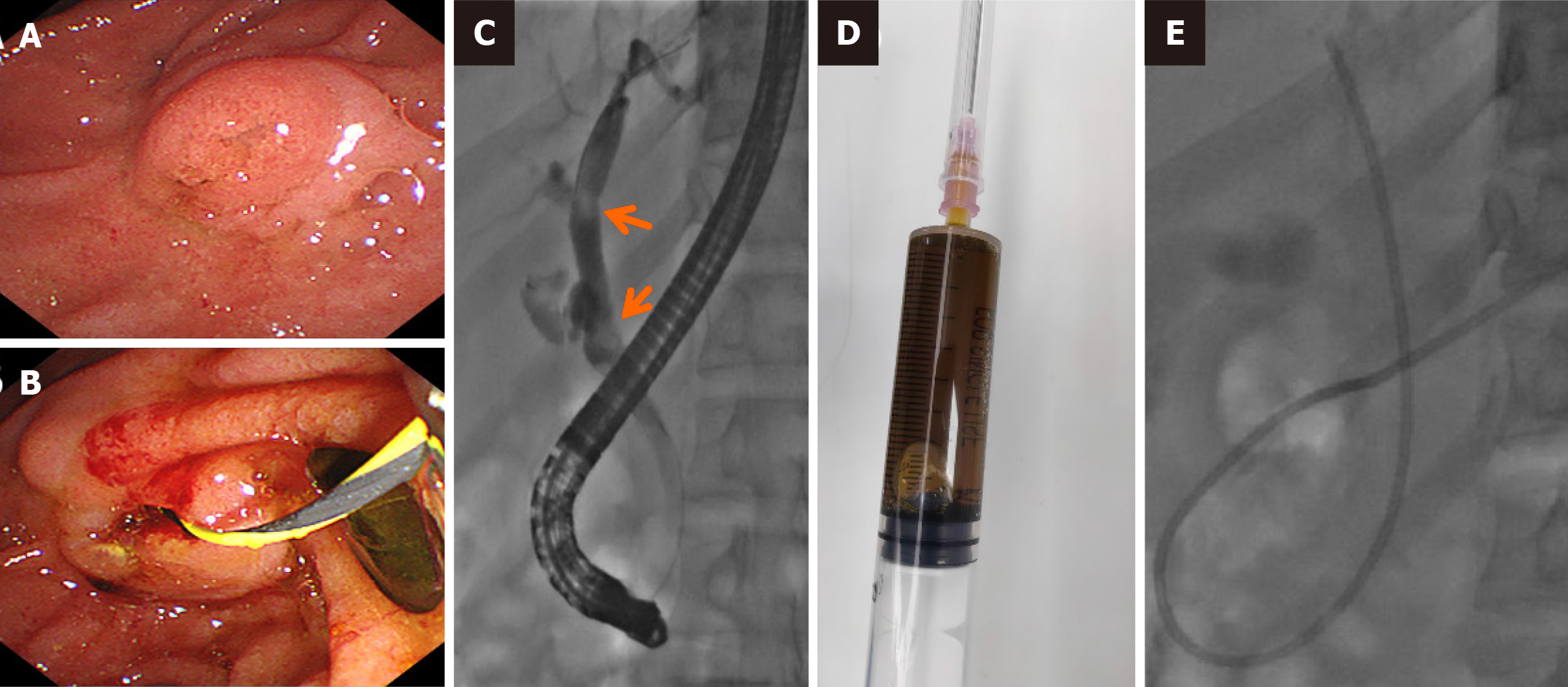
Figure 4 Two openings were detected in the duodenal papilla, revealing the distinct connection of the pancreatic duct and biliary duct to the intestinal wall.
A: Duodenal papilla; B: Black stone dragged into the duodenal cavity; C: Gallstones (arrow) in the common bile duct during endoscopic retrograde cholangiopancreatography; D: Bile fluid extracted during endoscopic retrograde cholangiopancreatography; E: Endoscopic nasobiliary drainage.
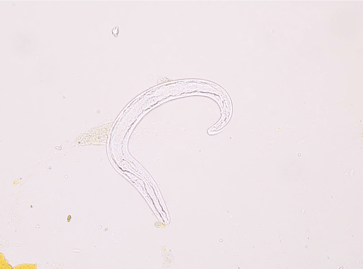
Figure 5 Alive Strongyloides stercoralis rhabditiform larvae discovered in bile fluid, as depicted under a microscope.
- Citation: Jiang XH, Deng Q, Wu ZK, Li JZ. Alive Strongyloides stercoralis in biliary fluid in patient: A case report. World J Gastroenterol 2025; 31(4): 98752
- URL: https://www.wjgnet.com/1007-9327/full/v31/i4/98752.htm
- DOI: https://dx.doi.org/10.3748/wjg.v31.i4.98752


