Copyright
©The Author(s) 2025.
World J Gastroenterol. Jan 28, 2025; 31(4): 101288
Published online Jan 28, 2025. doi: 10.3748/wjg.v31.i4.101288
Published online Jan 28, 2025. doi: 10.3748/wjg.v31.i4.101288
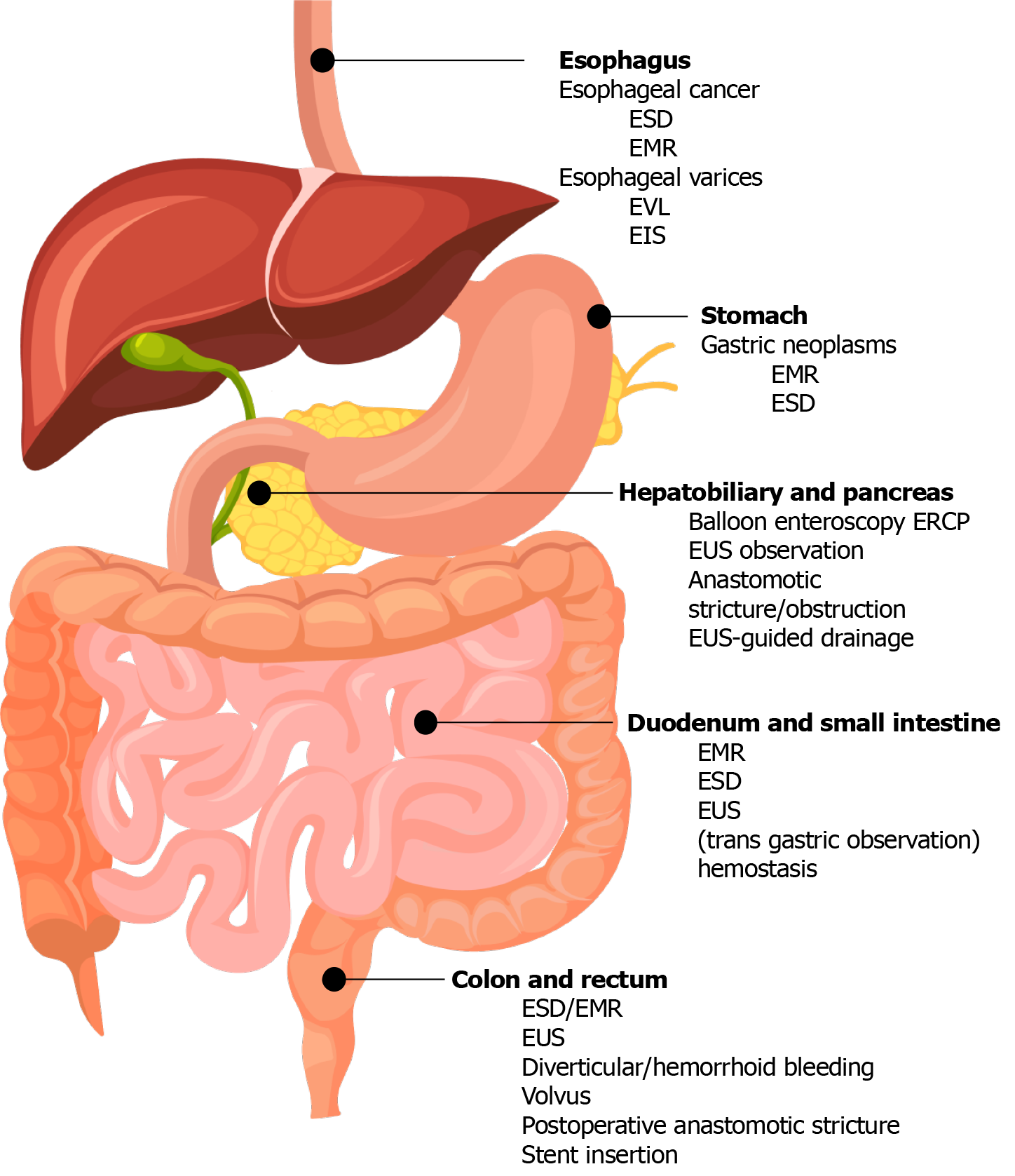
Figure 1 Gel use in gastrointestinal endoscopy is widely applied in all gastrointestinal tracts.
It is particularly characterized by its application in tumor resection and hemostasis in the stomach, intestine, and colon; esophageal varices treatment in the esophagus; and endoscopic ultrasound procedures in the hepatobiliary and pancreatic regions. ESD: Endoscopic submucosal dissection; EMR: Endoscopic mucosal resection; EUS: Endoscopic ultrasound; EVL: Endoscopic variceal ligation; EIS: Endoscopic injection sclerotherapy; ERCP: Endoscopic retrograde cholangiopancreatography. The authors have obtained the permission for figure using from the Adobe Stockphoto (Supplementary material).
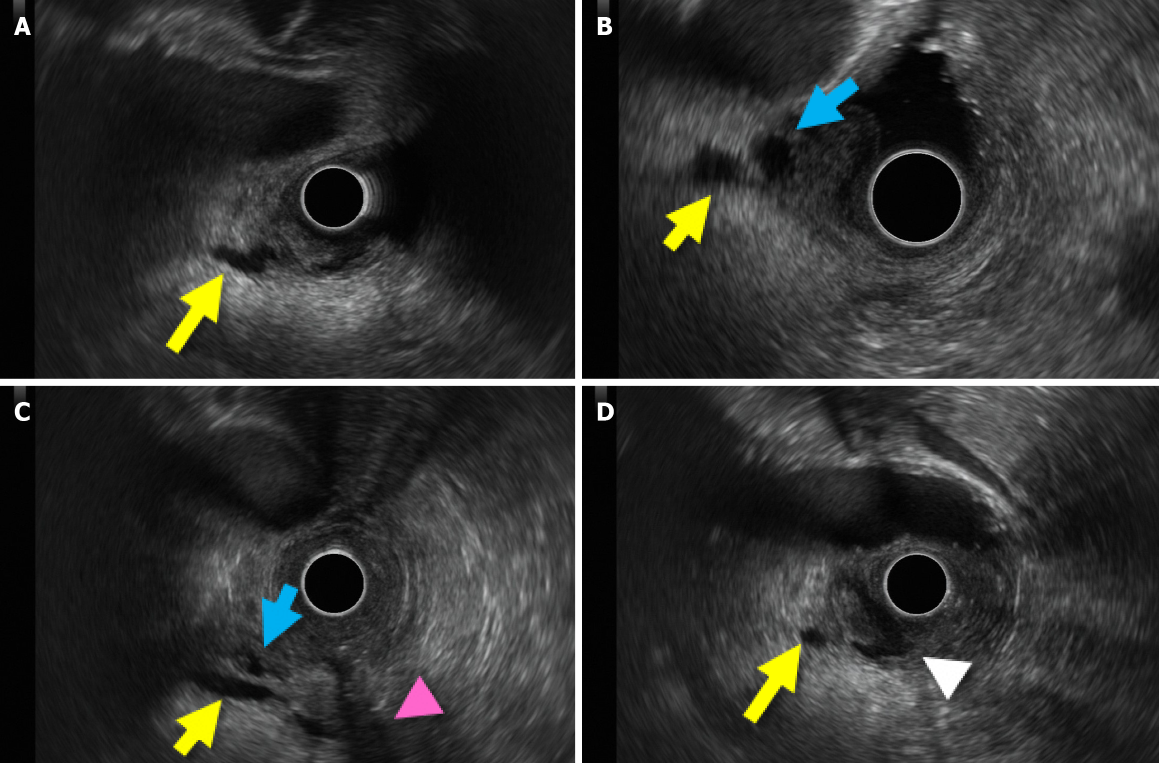
Figure 2 Utility of gel in endoscopic ultrasound for hepatobiliary and pancreatic visualization.
A: Clear depiction of the main pancreatic duct near the papilla (yellow arrow); B: The confluence of the main pancreatic duct and bile duct (blue arrow); C: Pancreatic and bile ducts relation to the duodenal muscle layer (magenta arrowhead); D: The pancreatic duct and the ampulla of Vater (white arrowhead) are depicted.
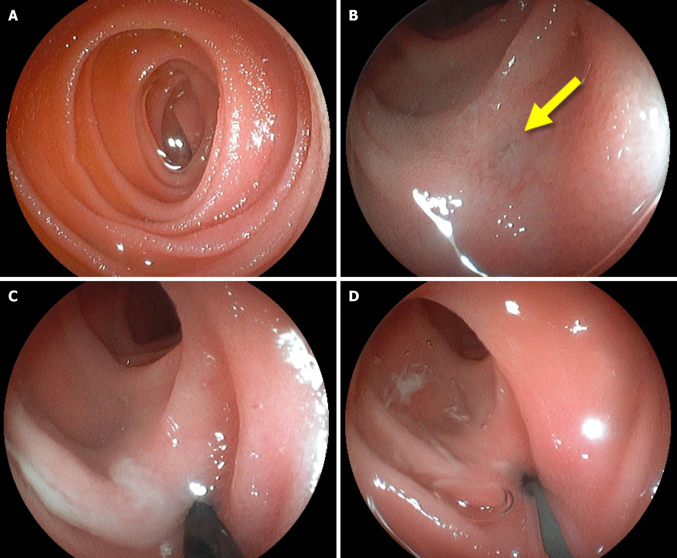
Figure 3 Utility of balloon-enteroscopy for hepatobiliary and pancreatic visualization.
A: The pancreaticojejunostomy opening is unclear without gel; B: Gel use enables identification of the pancreaticojejunostomy (yellow arrow); C: Catheter insertion through the identified pancreaticojejunostomy is achieved; D: The same site is dilated with a balloon catheter.
- Citation: Sato H, Kawabata H, Fujiya M. Gel immersion in endoscopy: Exploring potential applications. World J Gastroenterol 2025; 31(4): 101288
- URL: https://www.wjgnet.com/1007-9327/full/v31/i4/101288.htm
- DOI: https://dx.doi.org/10.3748/wjg.v31.i4.101288


