Copyright
©The Author(s) 2025.
World J Gastroenterol. May 14, 2025; 31(18): 105530
Published online May 14, 2025. doi: 10.3748/wjg.v31.i18.105530
Published online May 14, 2025. doi: 10.3748/wjg.v31.i18.105530
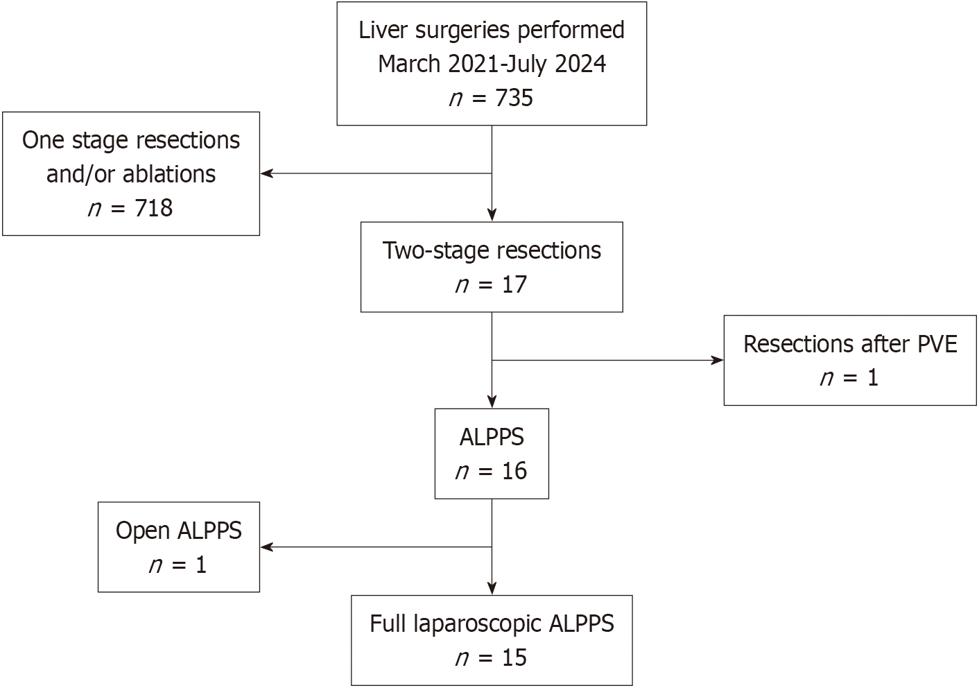
Figure 1 The flowchart shows all consecutive patients with colorectal liver metastases who underwent liver surgery at our institution between March 2021 and July 2024.
PVE: Portal venous embolization; ALPPS: Associating liver partition and portal vein ligation for staged hepatectomy.
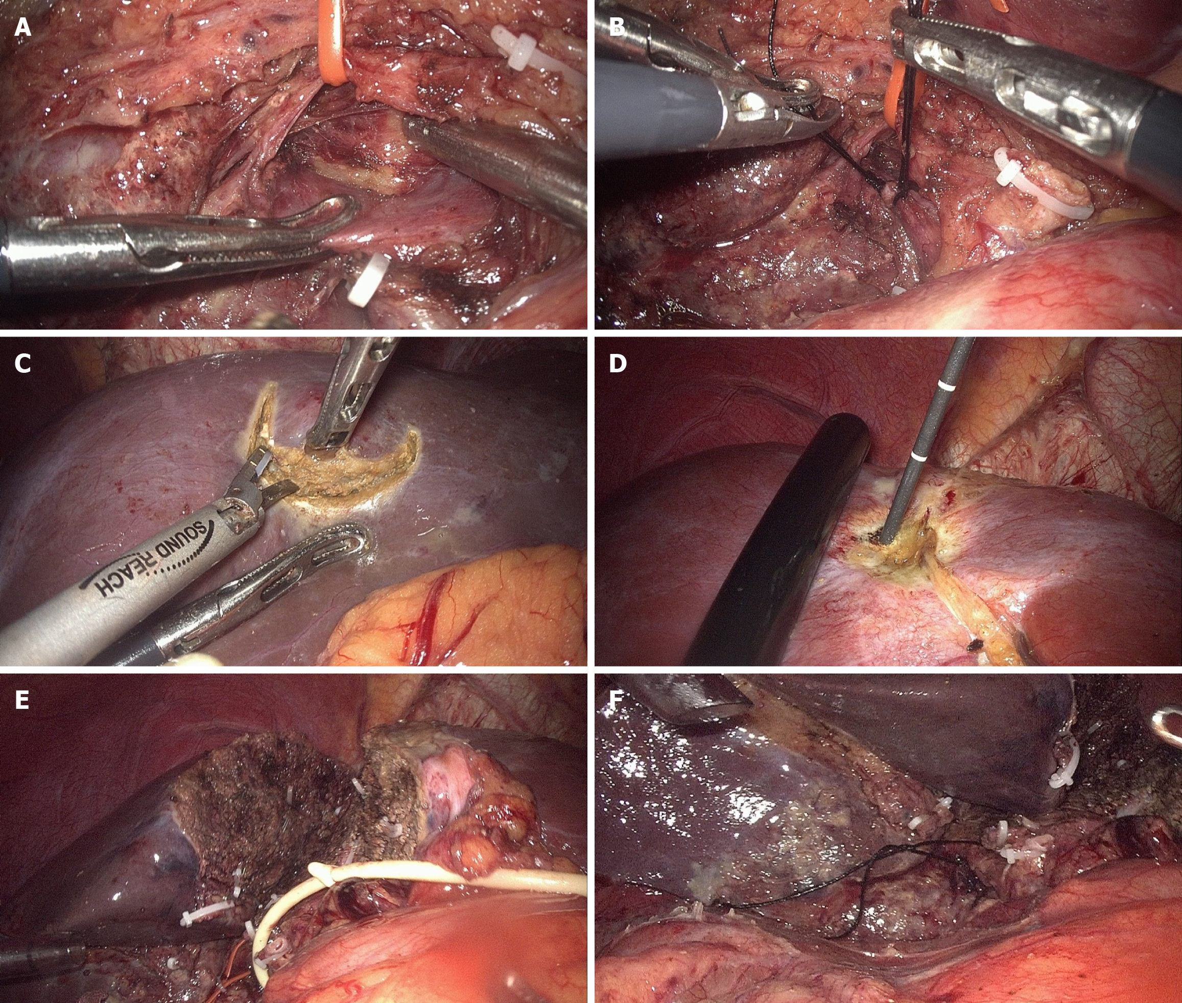
Figure 2 Main procedures in stage 1 of full laparoscopic associating liver partition and portal vein ligation for staged hepatectomy.
A: Identification of the right hepatic artery (RHA) and right portal vein with the RHA preserved using a tie; B: Ligation of right portal vein by double non-absorbable sutures; C: Radical resection of tumor in future liver remnant; D: Microwave ablation of tumor in future liver remnant; E: Partial transection of liver parenchyma in situ; F: Marking of the RHA is by a loose suture for identification during stage 2.
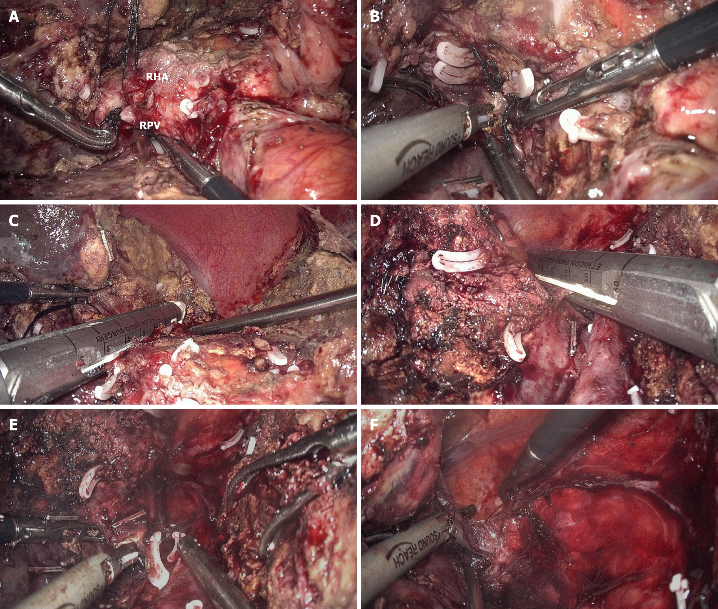
Figure 3 Main procedures in stage 2 of full laparoscopic associating liver partition and portal vein ligation for staged hepatectomy.
A: Identification of the right hepatic artery (the single long loose suture) and right portal vein (the double short tight sutures); B: After transection of the right hepatic artery and right portal vein, dissection of the space posterior to the right Glisson pedicle is taken; C: Division of the right Glisson pedicle by endostapler; D: Transection of the right hepatic vein by endostapler; E and F: Completely mobilization of the right liver as the final step of stage 2. RHA: Right hepatic artery; RPV: Right portal vein.
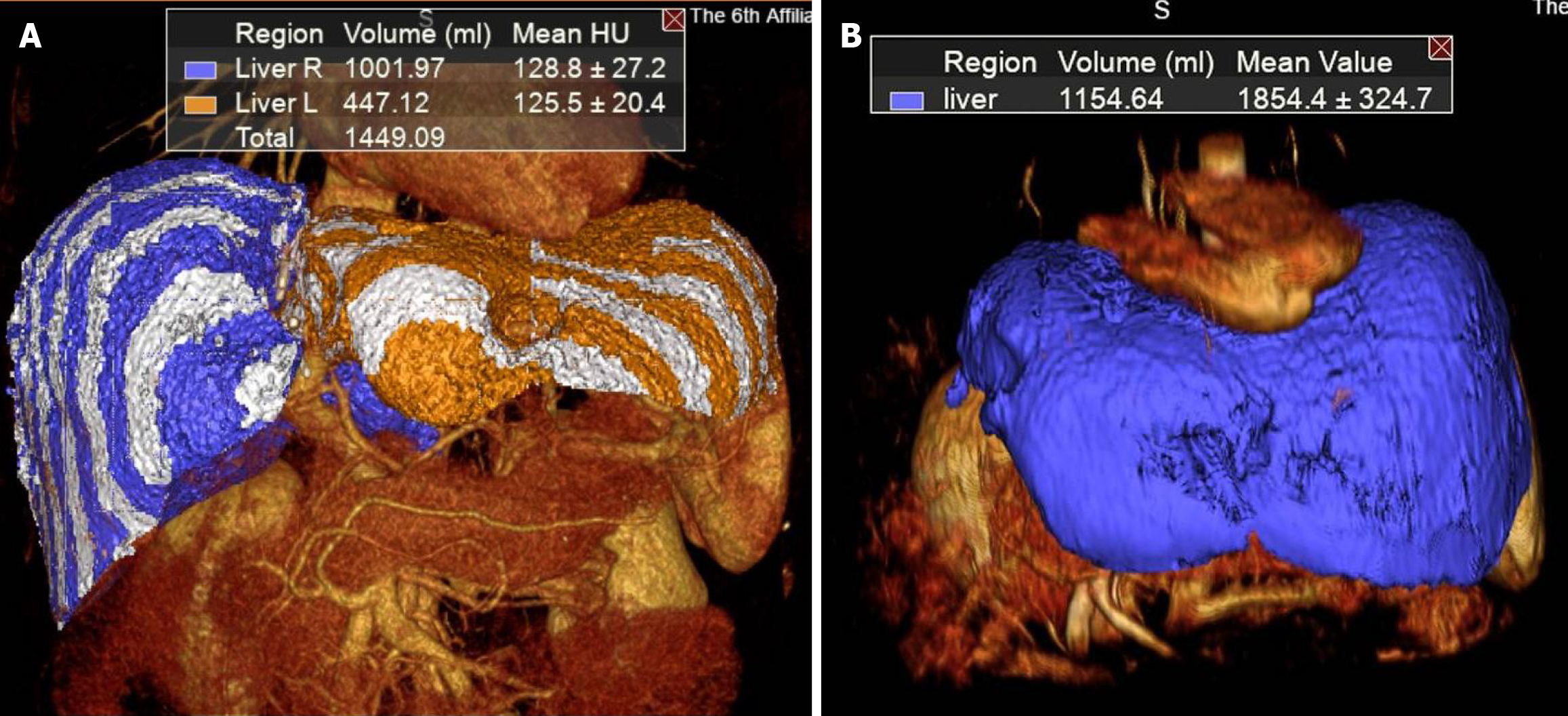
Figure 4 Regeneration of future liver remnant in the period of post-associating liver partition and portal vein ligation for staged hepatectomy.
A: Future liver remnant volume before stage 2 (447.1 mL); B: Future liver remnant volume at 3 months after stage 2 (1154.6 mL).
- Citation: Zheng ZY, Zhang L, Li WL, Dong SY, Song JL, Zhang DW, Huang XM, Pan WD. Laparoscopic associating liver partition and portal vein ligation for staged hepatectomy for colorectal liver metastases: A single-center experience. World J Gastroenterol 2025; 31(18): 105530
- URL: https://www.wjgnet.com/1007-9327/full/v31/i18/105530.htm
- DOI: https://dx.doi.org/10.3748/wjg.v31.i18.105530


