Copyright
©The Author(s) 2025.
World J Gastroenterol. May 7, 2025; 31(17): 105347
Published online May 7, 2025. doi: 10.3748/wjg.v31.i17.105347
Published online May 7, 2025. doi: 10.3748/wjg.v31.i17.105347
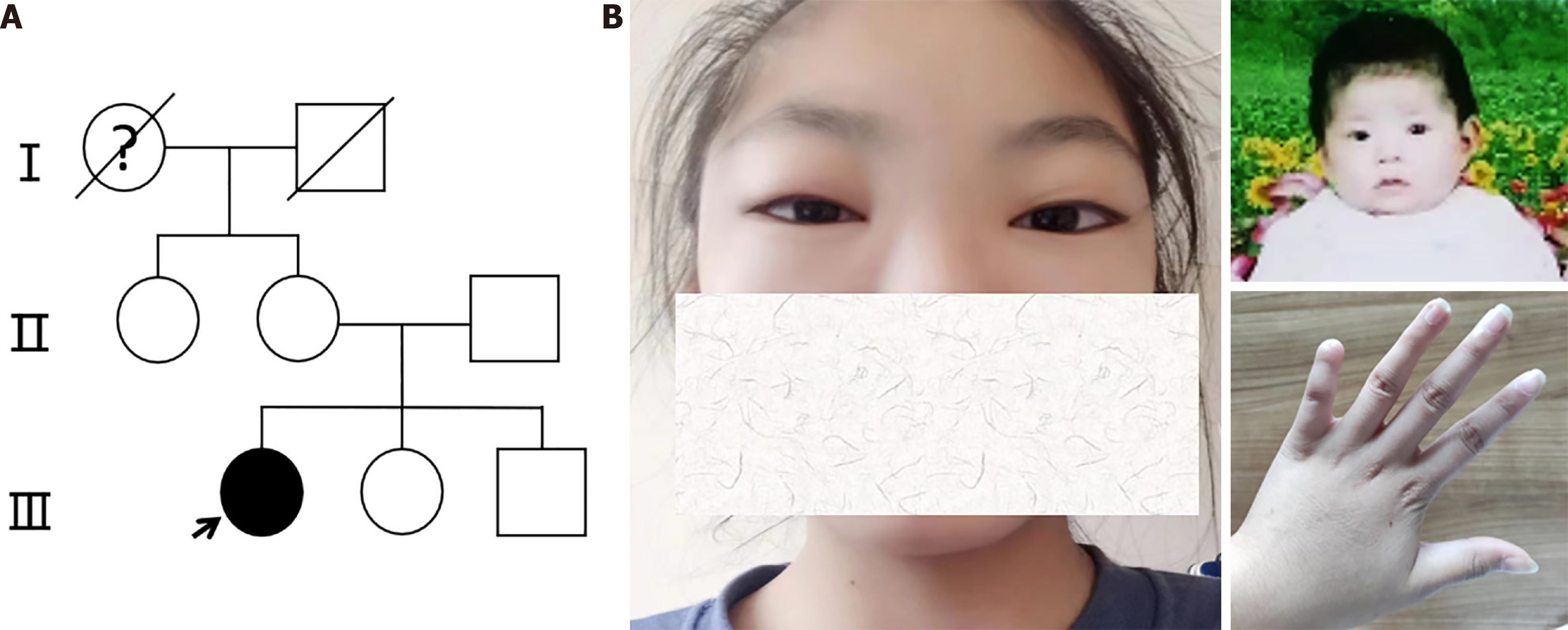
Figure 1 Pedigree and characteristic appearance of the patient.
A: In the pedigree of the patient’s family, patients are represented in black, the arrow represents the proband, which is the patient discussed in this case; B: Characteristic craniofacial appearance of widely spaced eyes in adolescence and infant. In addition, the distal phalanx of the left little finger was absent since birth. The patient was informed and agreed to publish the photos (Supplementary material).
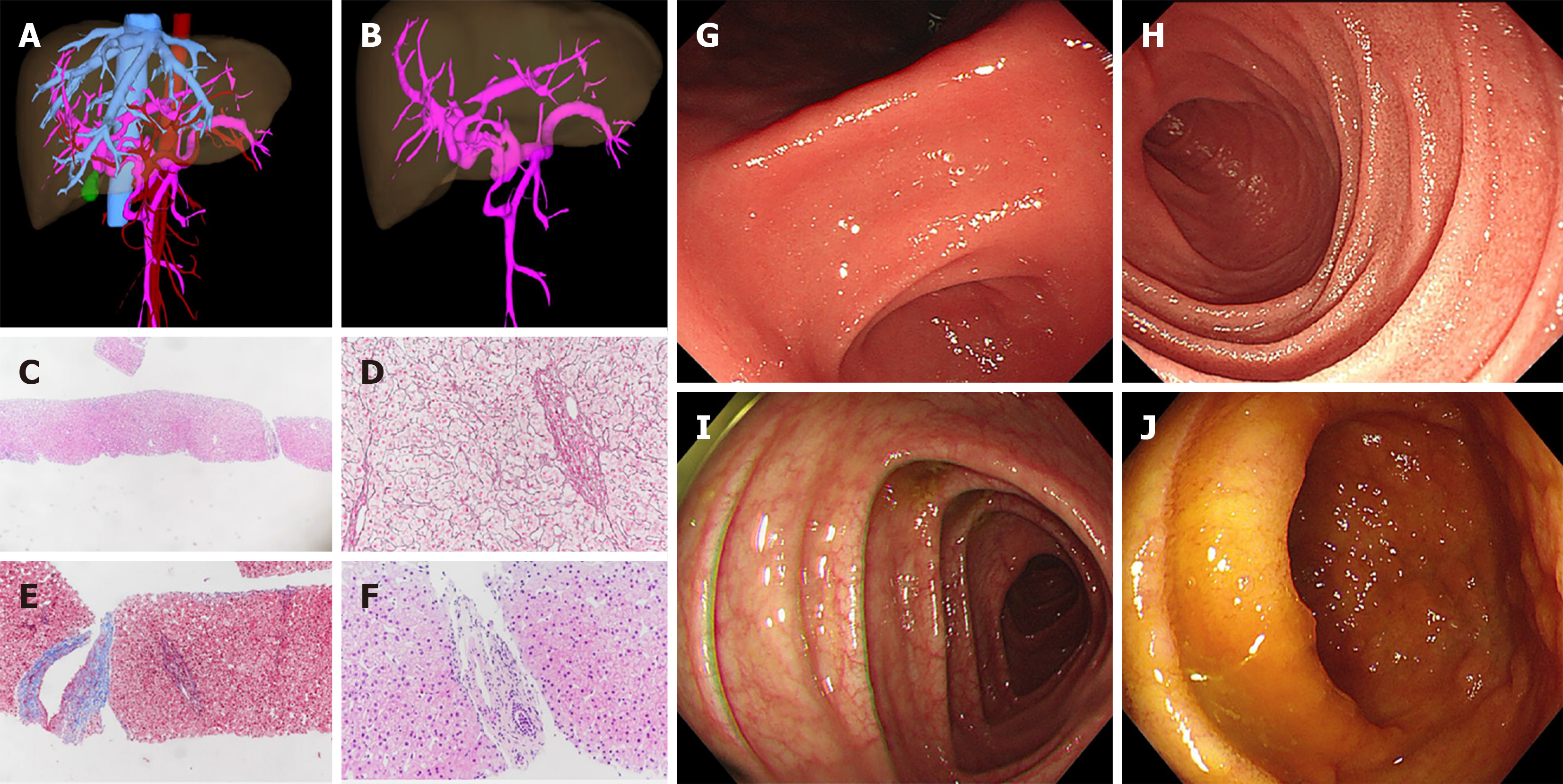
Figure 2 Imaging presentation about the liver and gastrointestinal tract.
A and B: Abdominal enhanced computed tomography by three-dimensional reconstruction indicating cavernous transformation of portal vein; C-F: Liver histology suggesting porto-sinusoidal vascular disease. The discernable lobular architecture (C: Hematoxylin and eosin staining, 200 ×), stenosis or disappearance of portal veins in portal areas (D: Reticular staining, 400 ×), herniated portal veins into the liver parenchyma, and smooth muscle proliferation in portal areas (E: Masson-trichrome staining, 200 ×), inflammatory cell infiltration not obvious in the portal areas (F: Hematoxylin and eosin staining, 400 ×); G and H: Upper and lower endoscopies showed no obvious abnormalities in the gastric and duodenal mucosa; I and J: No obvious abnormalities in the ileal and colonic mucosa except for the slight edema of the colon wall.
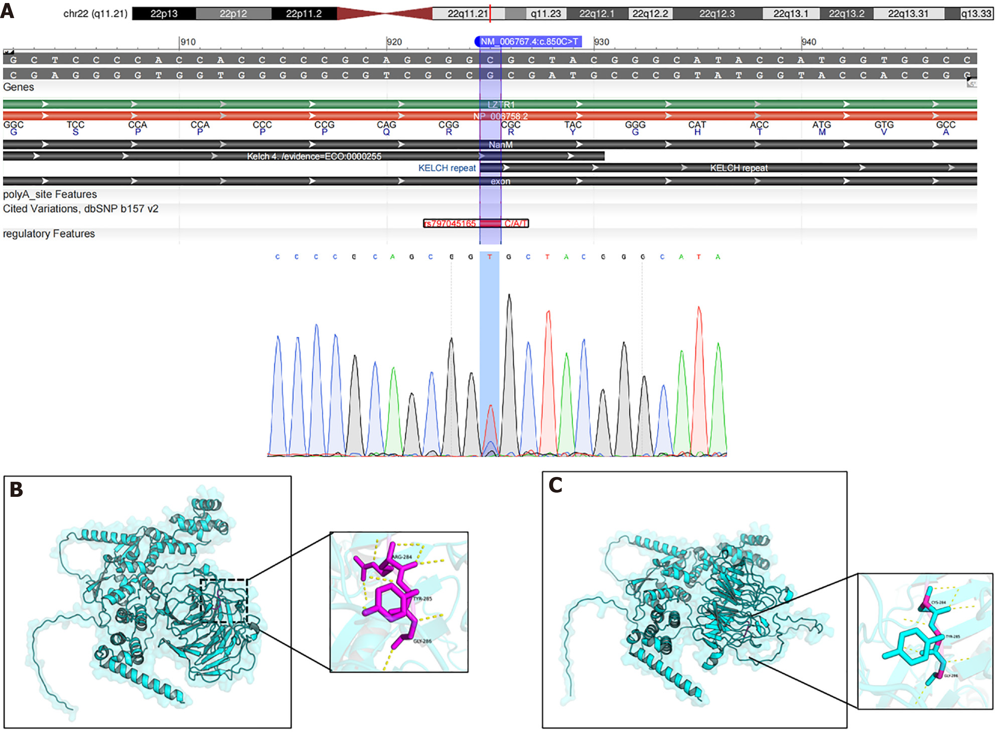
Figure 3 Pathogenic genetic variance information of the patient.
A: Whole-exome and Sanger sequencing revealed a heterozygous mutation (c.850C>T:p.Arg284Cys) in leucine zipper-like transcription regulator 1; B: Visualization of the protein structures of wild-type using Pymol software; C: Visualization of the protein structures of mutant (c.850C>T:P.R284C) leucine zipper-like transcription regulator 1. After the mutation, the amino acid residue at position 284 changes from arginine to cysteine, resulting in a decrease in the number of hydrogen bonds, which may lead to a loosening of the local structure (the yellow dashed lines representing hydrogen bonds).
- Citation: Tian QJ, Zhang LJ, Zhang Q, Liu FC, Xie M, Cai JZ, Rao W. Protein-losing enteropathy and multiple vasculature dysplasia in LZTR1-related Noonan syndrome: A case report and review of literature. World J Gastroenterol 2025; 31(17): 105347
- URL: https://www.wjgnet.com/1007-9327/full/v31/i17/105347.htm
- DOI: https://dx.doi.org/10.3748/wjg.v31.i17.105347


