Copyright
©The Author(s) 2024.
World J Gastroenterol. Feb 7, 2024; 30(5): 462-470
Published online Feb 7, 2024. doi: 10.3748/wjg.v30.i5.462
Published online Feb 7, 2024. doi: 10.3748/wjg.v30.i5.462
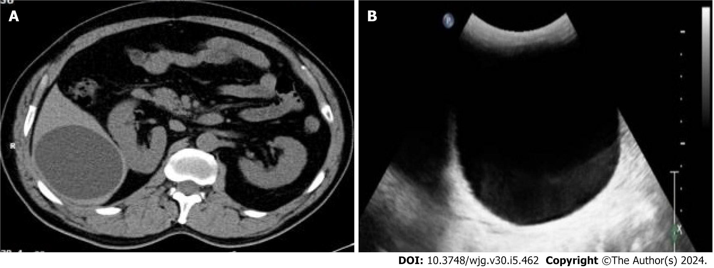
Figure 1 Hepatic cyst diagnosis.
A: Cystic echinococcosis type 1 (CE1) lesion, which was diagnosed as hepatic cyst by computed tomography; B: CE1 lesion, which was diagnosed as hepatic cyst by ultrasound.
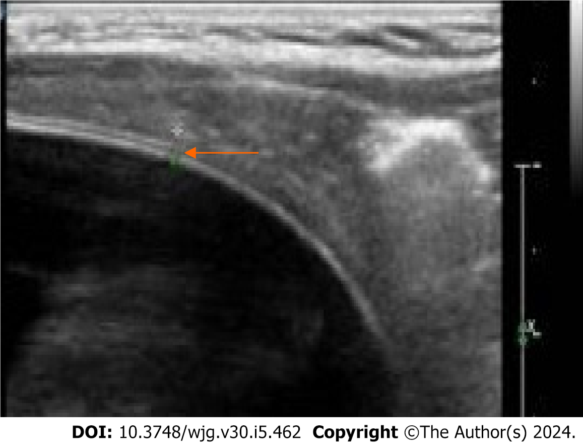
Figure 2 Hydatid cyst with a localized bilayered wall (arrowhead) detected by a linear array transducer.
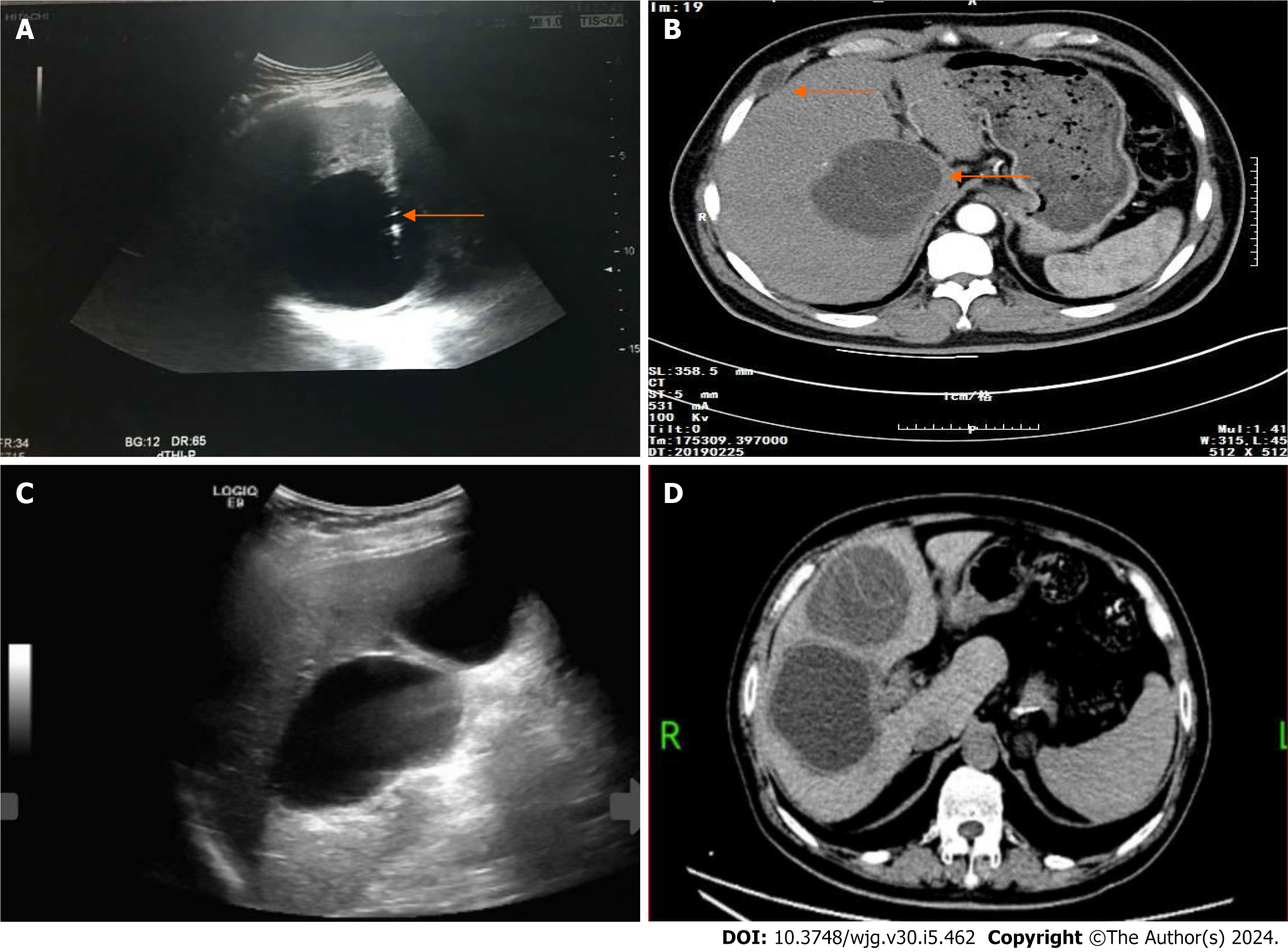
Figure 3 Hepatic cyst diagnosis and treatment.
A: Cystic echinococcosis type 1 (CE1) misdiagnosed as a hepatic cyst and treated by percutaneous aspiration, the arrowhead indicating the aspiration needle; B: Post-treatment follow-up revealing recurrence of a hepatic hydatid cyst (arrowhead) with hydatid implanted in the intercostal space (arrowhead); C: Conventional ultrasound diagnosing two simple hepatic cysts; D: Follow-up examination confirming the diagnosis of CE compressing the right hepatic pedicle.
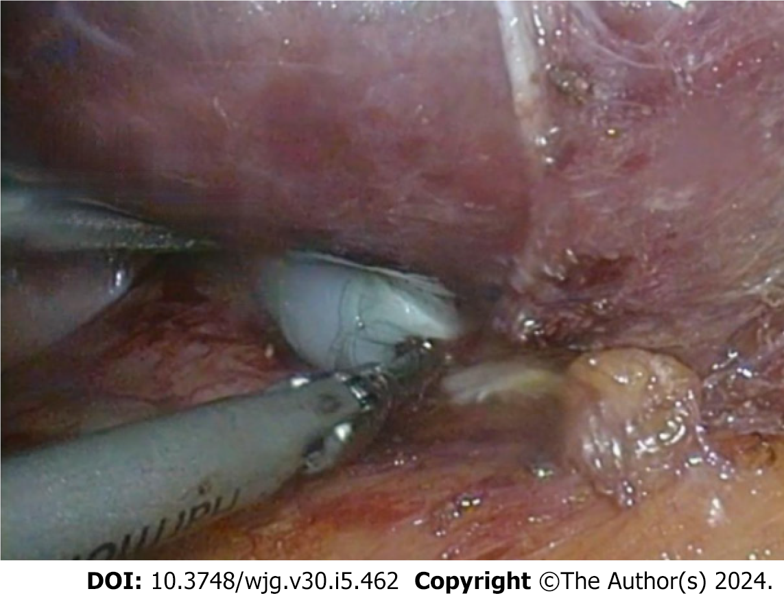
Figure 4 Endocyst of cystic echinococcosis type 1 under the laparoscope.
- Citation: Li YP, Zhang J, Li ZD, Ma C, Tian GL, Meng Y, Chen X, Ma ZG. Diagnosis and treatment experience of atypical hepatic cystic echinococcosis type 1 at a tertiary center in China. World J Gastroenterol 2024; 30(5): 462-470
- URL: https://www.wjgnet.com/1007-9327/full/v30/i5/462.htm
- DOI: https://dx.doi.org/10.3748/wjg.v30.i5.462


