Copyright
©The Author(s) 2024.
World J Gastroenterol. Nov 14, 2024; 30(42): 4557-4565
Published online Nov 14, 2024. doi: 10.3748/wjg.v30.i42.4557
Published online Nov 14, 2024. doi: 10.3748/wjg.v30.i42.4557
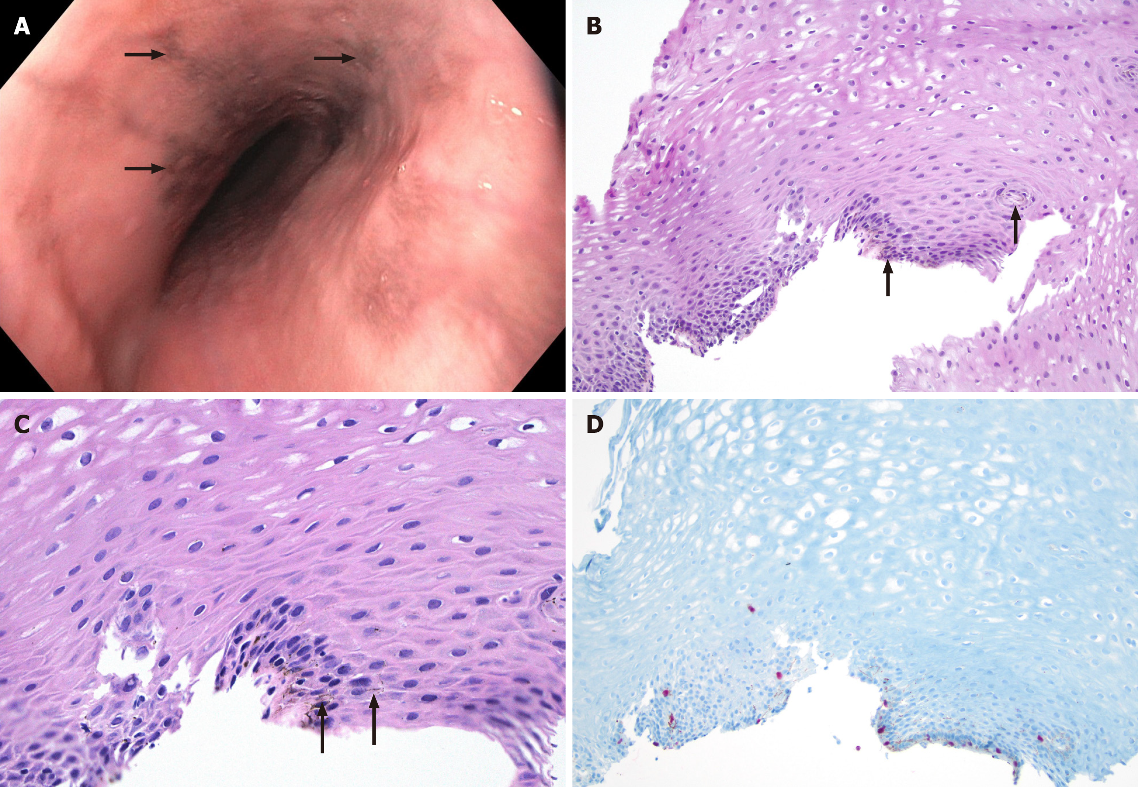
Figure 1 Imaging examinations.
A: Endoscopic poorly delineated pigmented lesion (arrow) on the mucosa; B and C: Hematoxylin and eosin stain shows basal cell hyperplasia, intercellular edema, and presence on non-atypical cells with melanin deposition (arrow) along the basal layer of the epithelium, 10 × and 40 × magnification; D: SRY-related HMG box 10 immunohistochemistry red nuclear positivity in the melanocytes.
- Citation: Kazacheuskaya L, Arora K. Esophageal melanosis: Two case reports and review of literature. World J Gastroenterol 2024; 30(42): 4557-4565
- URL: https://www.wjgnet.com/1007-9327/full/v30/i42/4557.htm
- DOI: https://dx.doi.org/10.3748/wjg.v30.i42.4557


