Copyright
©The Author(s) 2024.
World J Gastroenterol. Nov 7, 2024; 30(41): 4439-4448
Published online Nov 7, 2024. doi: 10.3748/wjg.v30.i41.4439
Published online Nov 7, 2024. doi: 10.3748/wjg.v30.i41.4439
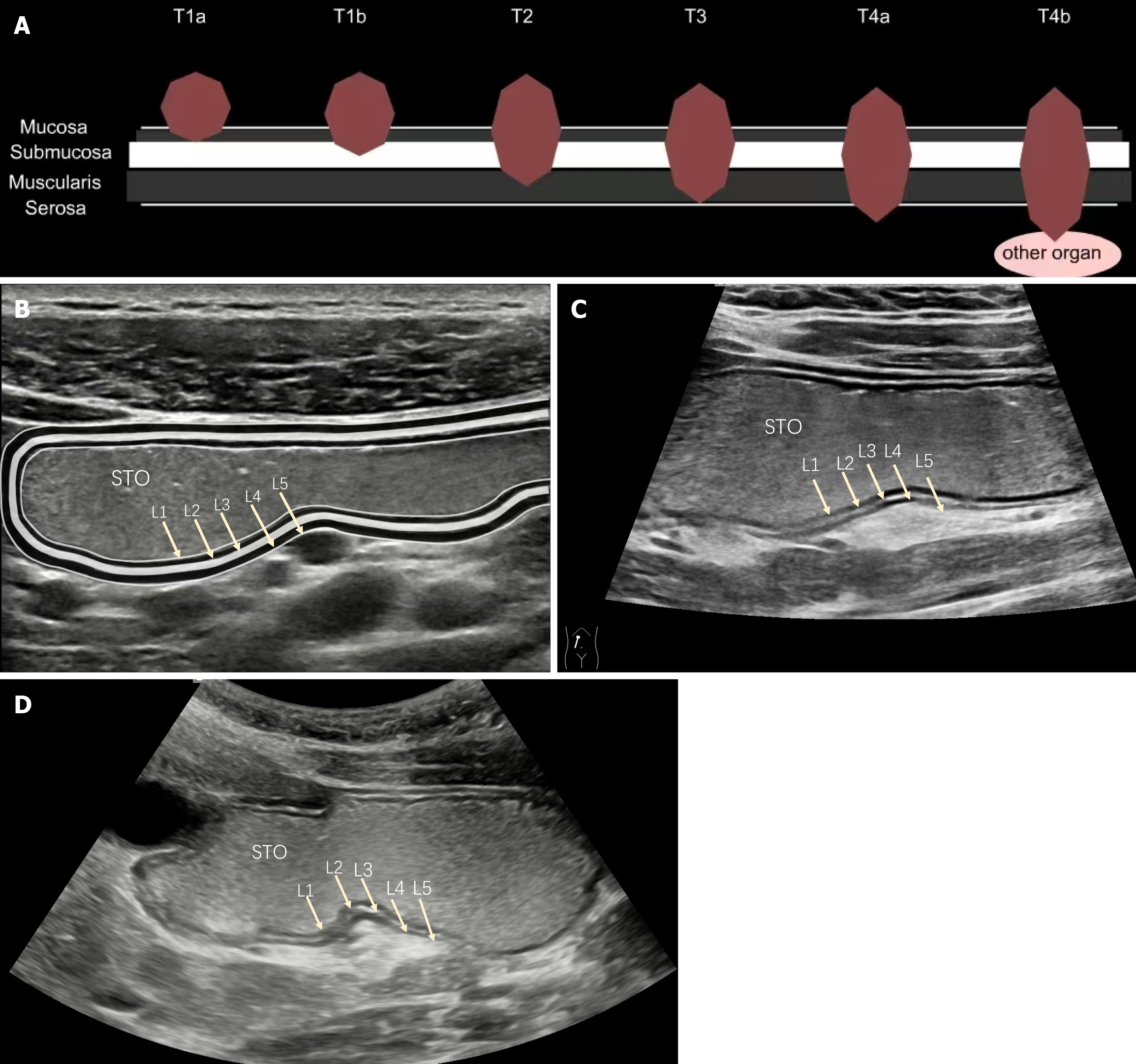
Figure 1 Oral contrast-enhanced ultrasound pattern chart of layers of the gastric wall.
A: Gastric cancer T staging pattern chart; B: Oral contrast-enhanced ultrasound pattern chart; C: Gastric oral contrast-enhanced ultrasound (OCEUS) high-frequency ultrasound pattern chart; D: Gastric OCEUS low-frequency ultrasound pattern chart. OCEUS reveals three high and two low five-layer structures of the gastric wall. L1: Mucosal epithelium and gastric cavity interface echo layer (high echo); L2: Deep mucosa (low echo); L3: Submucosa (high echo); L4: Muscularis propria (low echo); L5: Serosa (high echo); STO: Stomach cavity (equal echo).
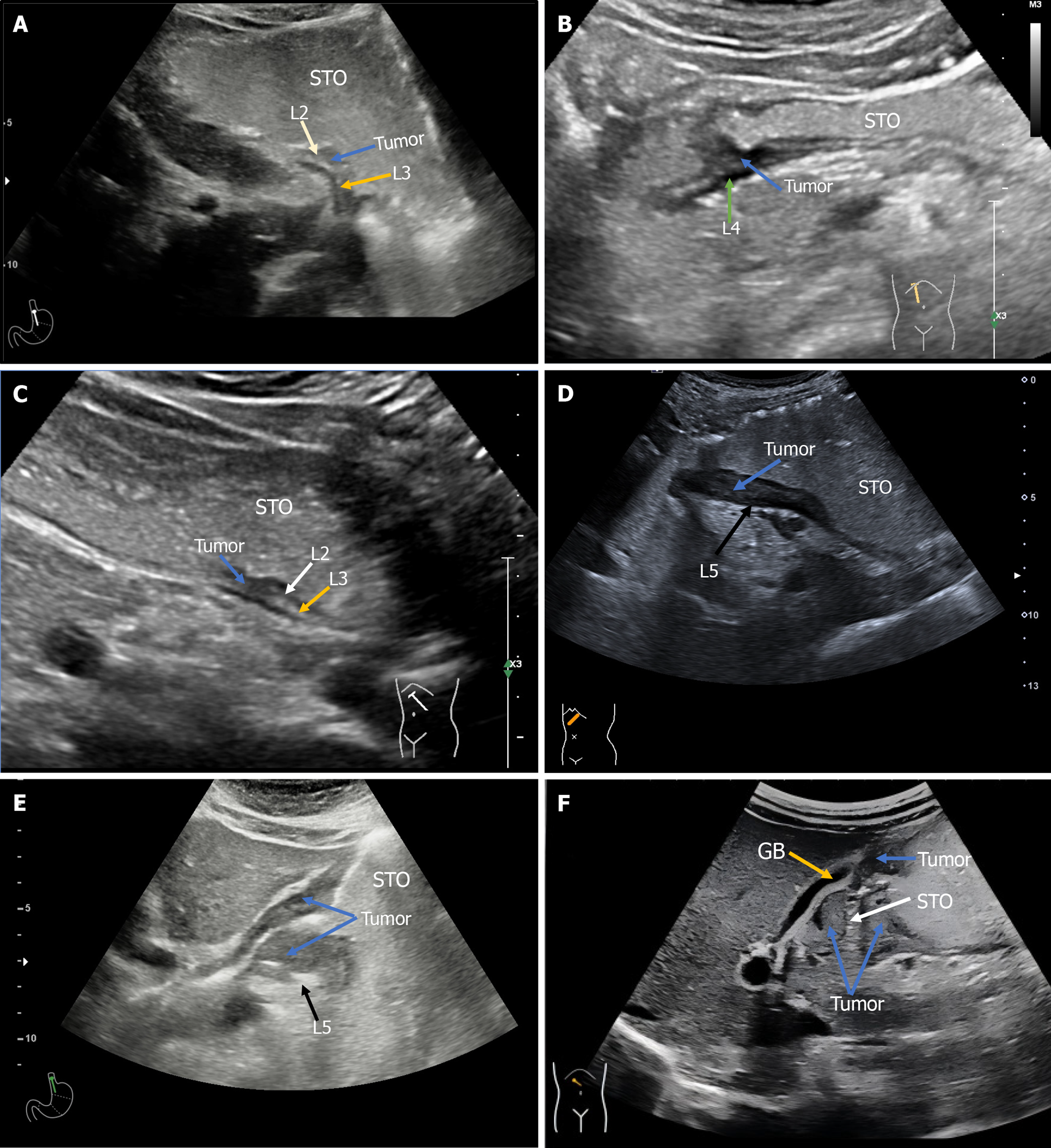
Figure 2 Case demonstration of each oral contrast-enhanced ultrasound T-stage.
A: Ultrasound T1a-stage; B: Ultrasound T1b-stage; C: Ultrasound T2-stage; D: Ultrasound T3-stage; E: Ultrasound T4a-stage; F: Ultrasound T4b-stage. L2: Deep mucosa; L3: Submucosa; L4: Muscularis propria; L5: Serosa; STO: Stomach cavity.
- Citation: Liang Y, Jing WY, Song J, Wei QX, Cai ZQ, Li J, Wu P, Wang D, Ma Y. Clinical application of oral contrast-enhanced ultrasound in evaluating the preoperative T staging of gastric cancer. World J Gastroenterol 2024; 30(41): 4439-4448
- URL: https://www.wjgnet.com/1007-9327/full/v30/i41/4439.htm
- DOI: https://dx.doi.org/10.3748/wjg.v30.i41.4439


