Copyright
©The Author(s) 2024.
World J Gastroenterol. Oct 7, 2024; 30(37): 4149-4155
Published online Oct 7, 2024. doi: 10.3748/wjg.v30.i37.4149
Published online Oct 7, 2024. doi: 10.3748/wjg.v30.i37.4149
Figure 1 Complete occlusion of the colorectal anastomosis.
A: Endoscopic view from the colonic side; B: 1% methylene blue solution was used to confirm the obstruction; C: Endoscopic view from the rectal side.
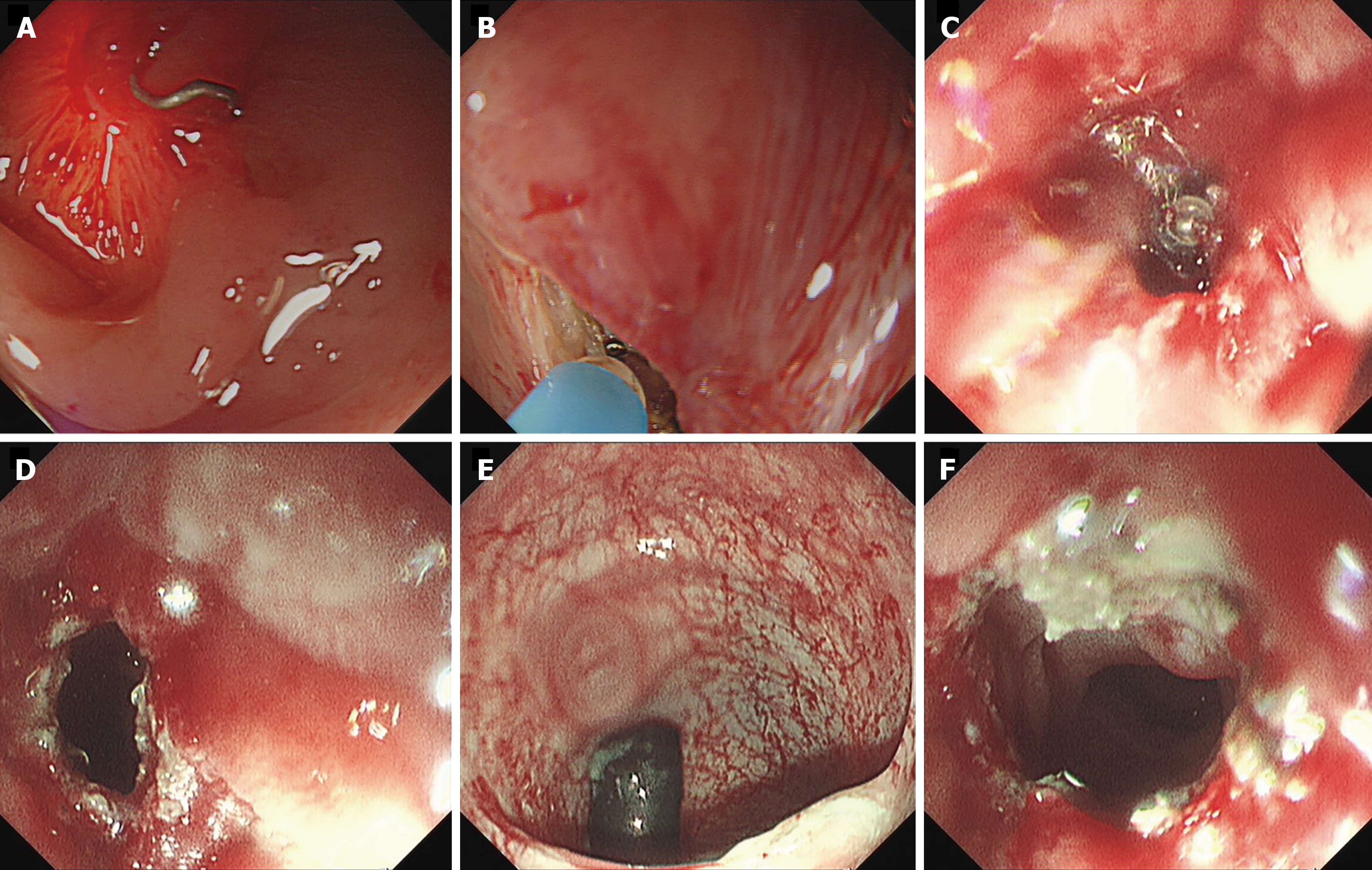
Figure 2 Details of endoscopic treatment.
A: Transillumination across the occlusive anastomosis; B: A small incision was made with an iKnife; C: Endoscopic view from the rectal side during the incision; D: Radial incision was performed successfully from the colonic side to the rectum; E: Endoscopic image showing dilation of the incision by the opposing endoscope; F: Recanalization of the occluded anastomosis.
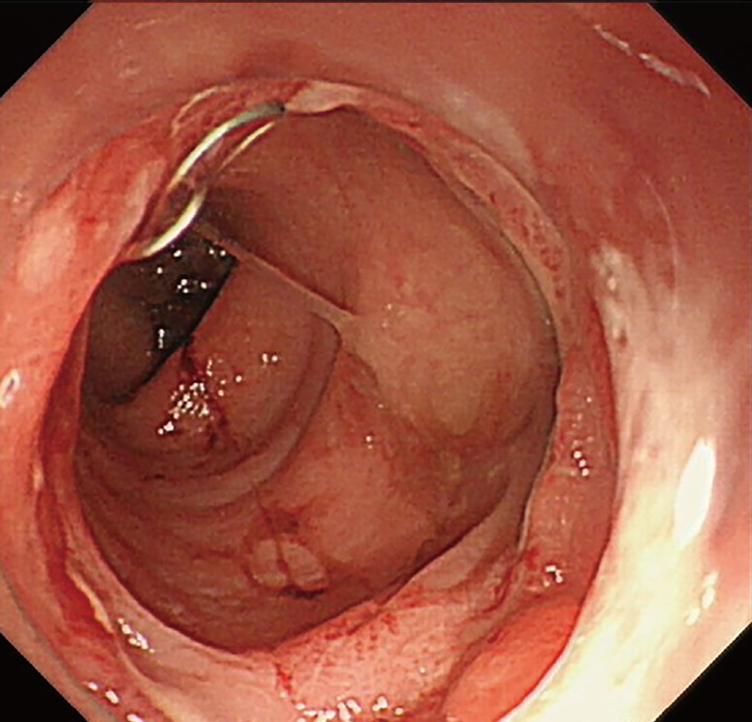
Figure 3 Endoscopy three weeks after treatment.
No anastomotic strictures or leakage were observed.
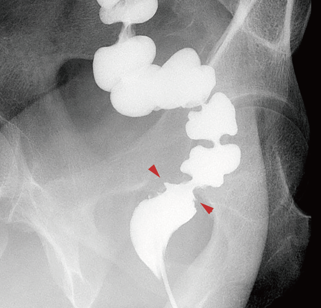
Figure 4 Air and barium double-contrast radiography.
Luminal continuity was observed to be re-established (arrows show the anastomosis).
- Citation: Chi J, Luo GY, Shan HB, Lin JZ, Wu XJ, Li JJ. Recanalization of anastomotic occlusion following rectal cancer surgery using a rendezvous endoscopic technique with transillumination: A case report. World J Gastroenterol 2024; 30(37): 4149-4155
- URL: https://www.wjgnet.com/1007-9327/full/v30/i37/4149.htm
- DOI: https://dx.doi.org/10.3748/wjg.v30.i37.4149


