Copyright
©The Author(s) 2024.
World J Gastroenterol. Oct 7, 2024; 30(37): 4115-4131
Published online Oct 7, 2024. doi: 10.3748/wjg.v30.i37.4115
Published online Oct 7, 2024. doi: 10.3748/wjg.v30.i37.4115
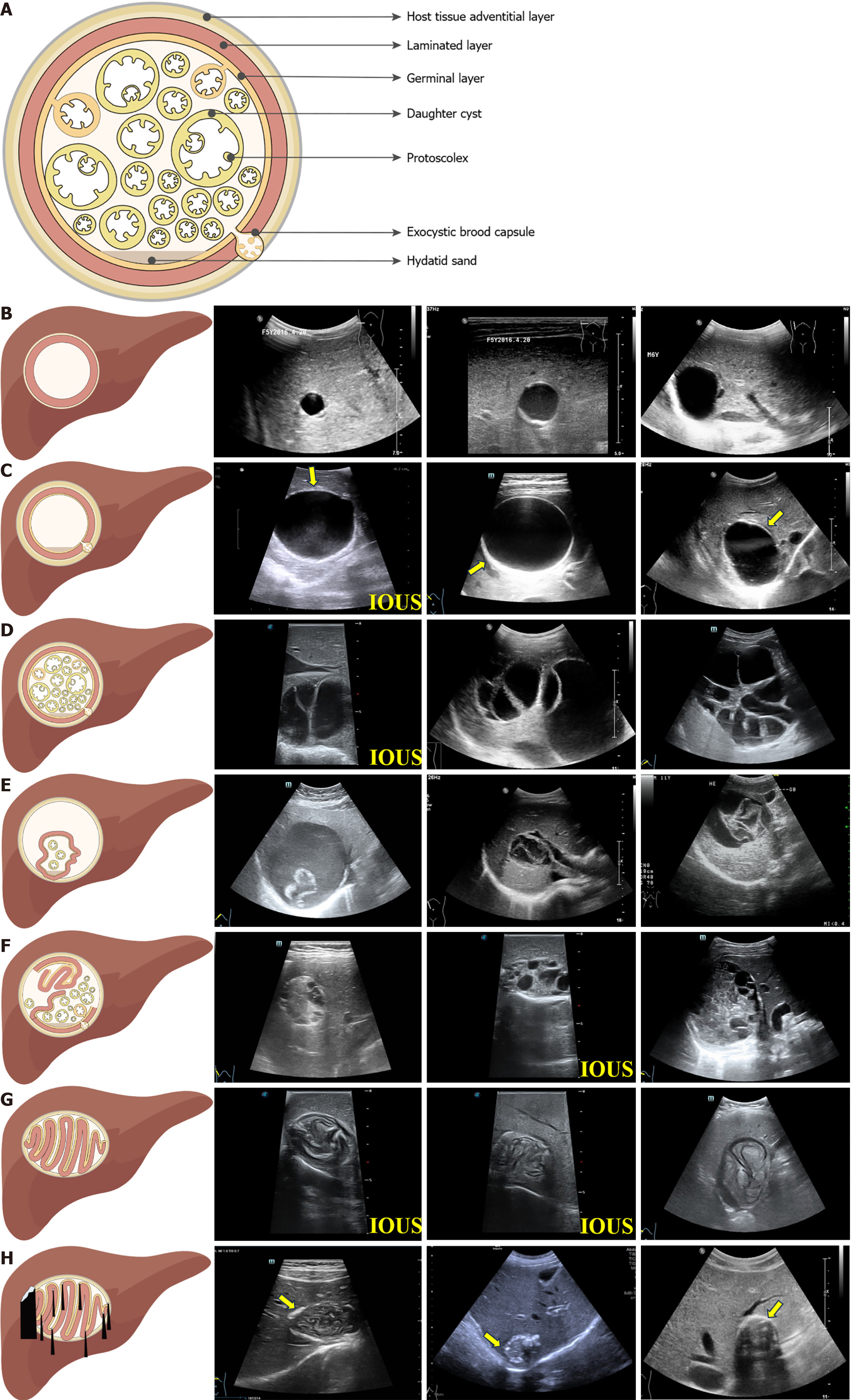
Figure 1 Schematic diagrams and grayscale ultrasound images of hepatic cystic echinococcosis.
A: Schematic structure of hepatic cystic echinococcosis (HCE); B: Hepatic cystic lesions: Hepatic cystic lesions are characterized by indistinct cyst walls, which can be more effectively visualized using high-frequency ultrasound transducers; C: HCE1: HCE1 lesions manifest as spherical, homogeneously echogenic cystic masses, characterized by the presence of “double cyst wall” (the yellow arrow)and “snowflake” signs; D: HCE2: HCE2 lesions are characterized by “nested cysts” or “daughter cysts” and wheel-, petal-, or honeycomb-like appearances; E: HCE3a: Features of HCE3a lesions include the “water lily” sign and “ribbon” sign; F: HCE3b: HCE3b lesions manifest as cystic and solid masses with mixed echogenicity; G: HCE4: HCE4 lesions present alternating hypoechoic and hyperechoic masses, characterized by “ball of wool” and “cerebral gyri” signs; H: HCE5: HCE5 lesions refer to hyperechoic patches on the basis of the “cerebral gyrus” sign, with thickened and calcified cyst walls accompanied by a broad posterior acoustic shadow, referred to as the “eggshell calcified wall” sign (the yellow arrow). IOUS: Intraoperative ultrasound.
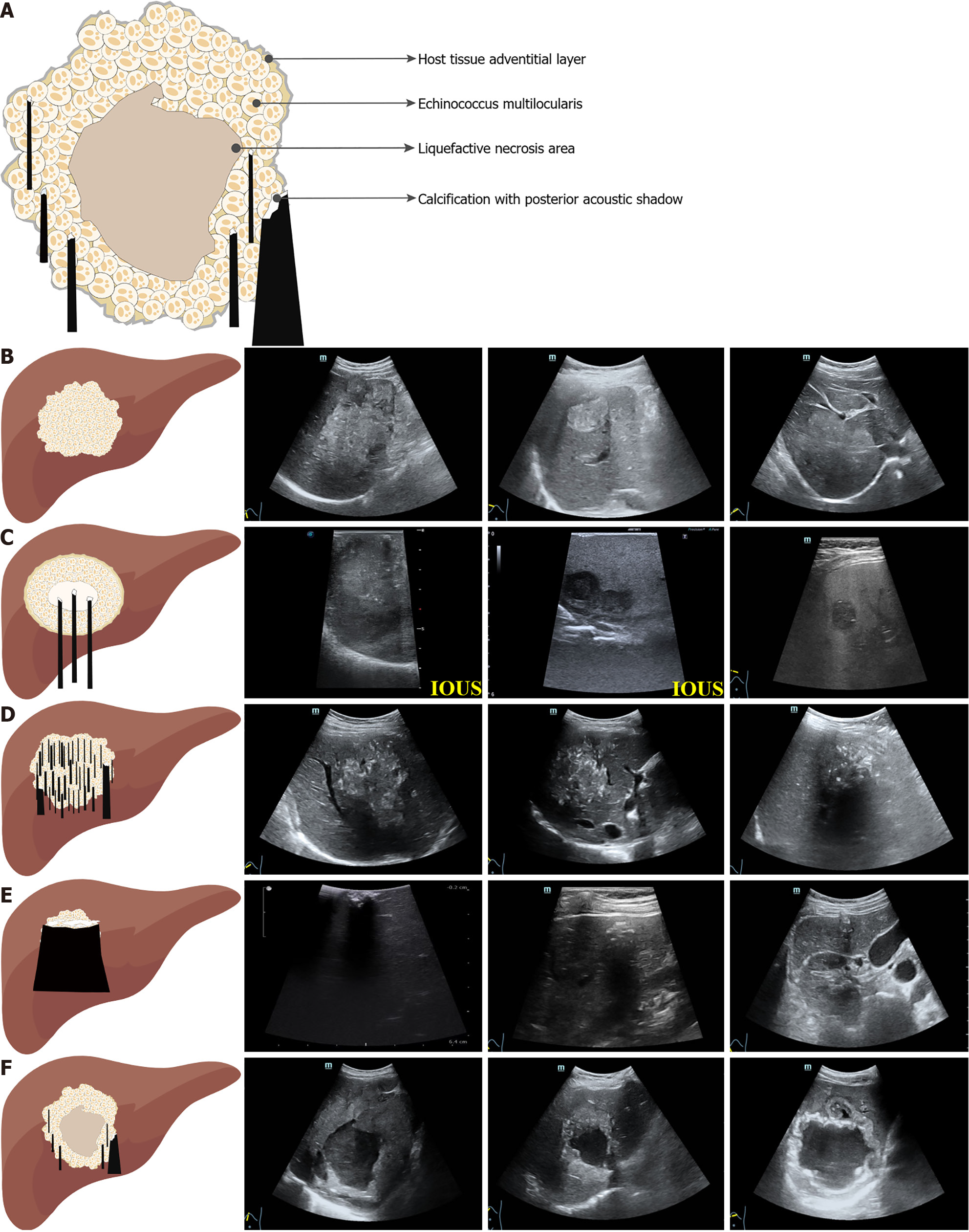
Figure 2 Schematic diagrams and grayscale ultrasound images of hepatic alveolar echinococcosis.
A: Schematic structure of hepatic alveolar echinococcosis (HAE); B: Hemangioma-like: Hemangioma-like lesions of HAE manifest as well-defined and heterogeneous hyperechoic masses in comparison to the surrounding hepatic parenchyma; C: Metastasis-like: Metastasis-like lesions of HAE manifest as hypoechoic masses with indistinct margins, lacking a halo sign, and with a hyperechoic and heterogeneous scar at their center; D: Hailstorm: Hailstorm lesions of HAE are characterized by ill-defined boundaries and irregularly shaped masses of heterogeneous echogenicity, accompanied by scattered or diffuse calcifications, with or without a posterior acoustic shadow; E: Ossification: Ossified lesions of HAE present as small and sharply delineated lesions with internal clumps of calcification and a posterior acoustic shadow; F: Pseudocystic: Pseudocystic lesions of HAE are primarily characterized by a thickness exceeding 10 mm with high echogenicity, irregular and non-uniform margins, and a low-echogenicity liquefied necrotic area inside. IOUS: Intraoperative ultrasound.
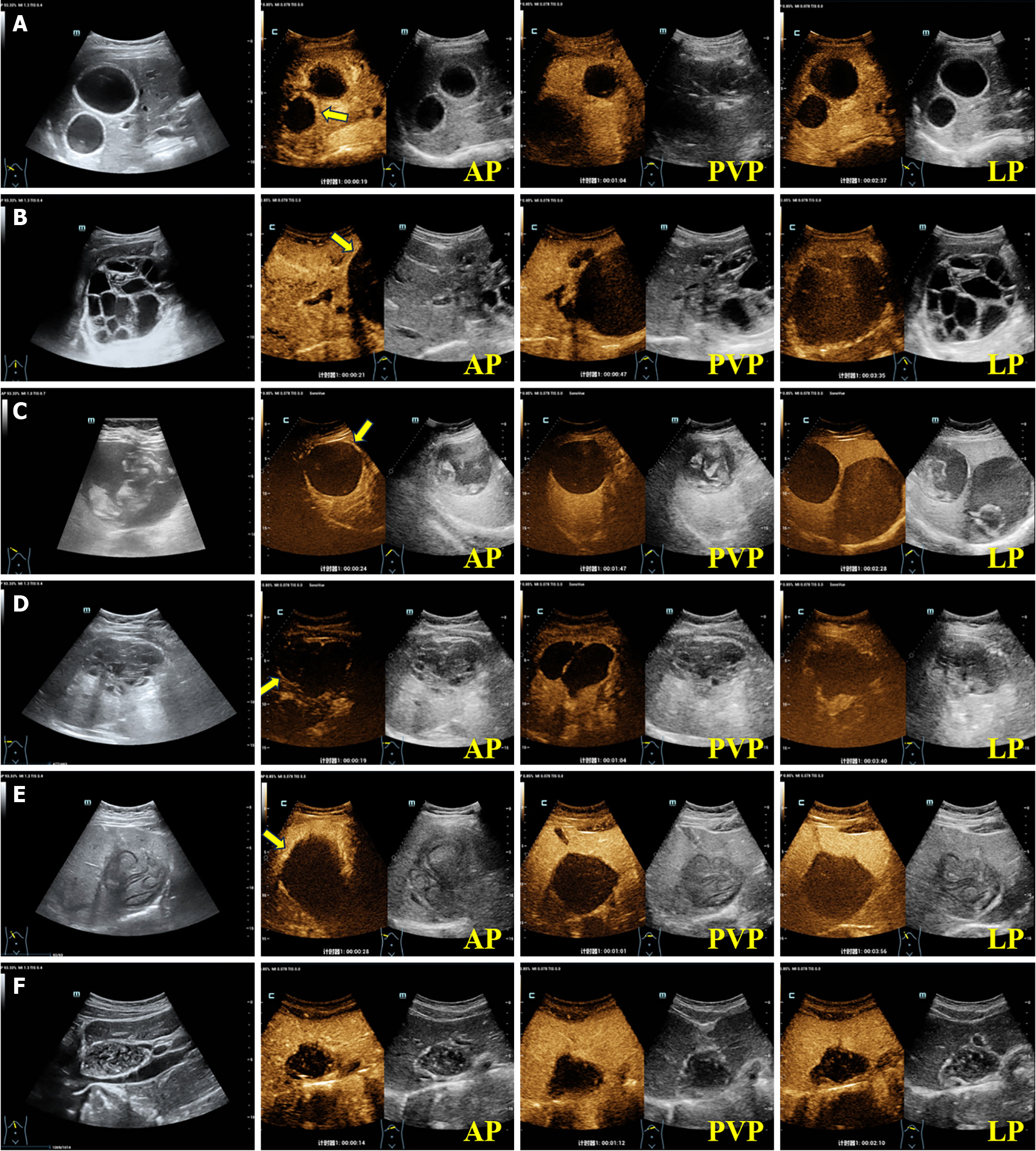
Figure 3 Contrast-enhanced ultrasound images of hepatic cystic echinococcosis.
A: A 27-year-old female patient presenting with a liver lesion of hepatic cystic echinococcosis 1 (HCE1), exhibiting peripheral “rim-like” enhancement in the arterial phase (AP) on contrast-enhanced ultrasound (CEUS) (the yellow arrow), with no internal enhancement observed; B: A 41-year-old female patient presenting with a liver lesion of HCE2, exhibiting slightly peripheral “rim-like” enhancement in the AP on CEUS (the yellow arrow), with no internal enhancement detected; C: A 56-year-old female patient presenting with a liver lesion of HCE3a, exhibiting slightly peripheral “rim-like” enhancement in the AP on CEUS (the yellow arrow), with no internal enhancement observed; D: A 49-year-old male patient presenting with a liver lesion of HCE3b, exhibiting slightly peripheral “rim-like” enhancement in the AP on CEUS (the yellow arrow), with internal septal enhancement visible; E: A 47-year-old female patient presenting with a liver lesion of HCE4, exhibiting slightly peripheral “rim-like” enhancement in the AP on CEUS (the yellow arrow), with no internal enhancement observed; F: A 29-year-old female patient presenting with a liver lesion of HCE5, with no obvious enhancement in the AP, portal venous phase, or late phase.
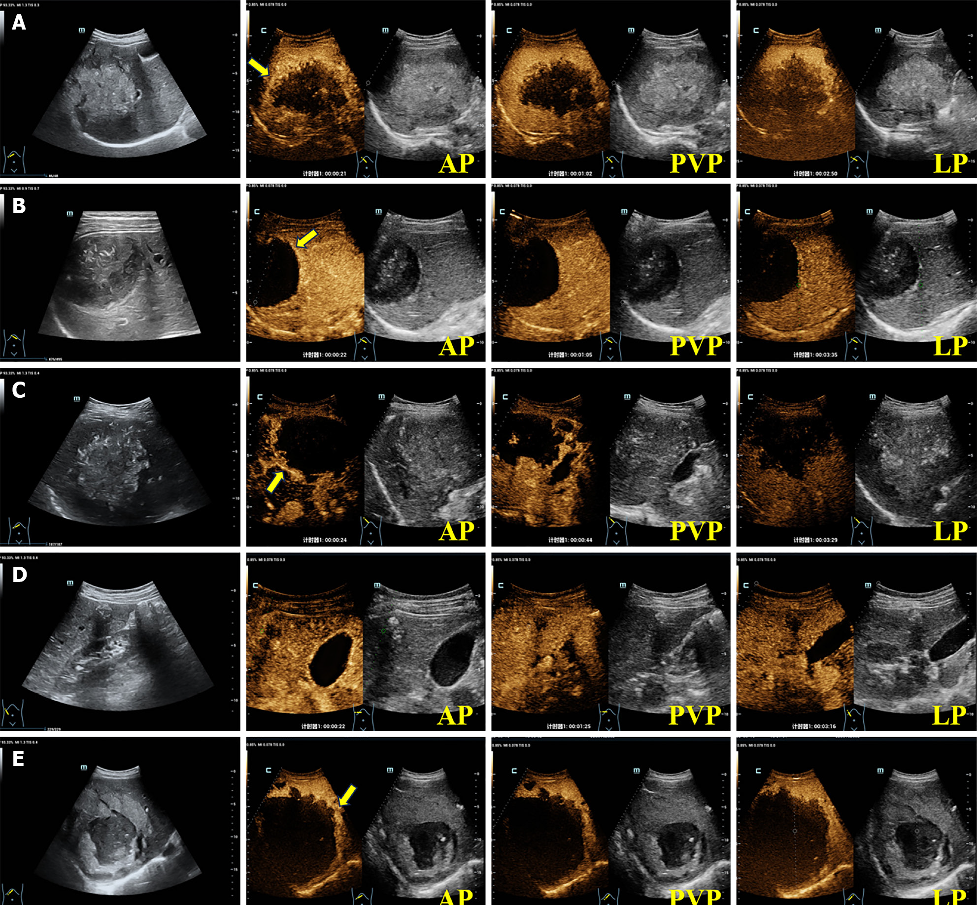
Figure 4 Contrast-enhanced ultrasound images of hepatic alveolar echinococcosis.
A: A 55-year-old male patient presenting with a hemangioma-like hepatic alveolar echinococcosis (HAE) liver lesion, exhibiting peripheral “rim-like” enhancement in the arterial phase (AP) on contrast-enhanced ultrasound (CEUS) (the yellow arrow), with no internal enhancement; B: A 29-year-old female patient presenting with a metastasis-like HAE liver lesion, exhibiting peripheral “rim-like” enhancement in the AP on CEUS (the yellow arrow), with no internal enhancement; C: A 53-year-old female patient presenting with a hailstorm HAE liver lesion, exhibiting peripheral “rim-like” enhancement in the AP on CEUS (the yellow arrow), with no internal enhancement; D: A 50-year-old male patient presenting with an ossified HAE liver lesion, with no obvious enhancement in the AP, portal venous phase, or late phase; E: A 40-year-old female patient presenting with a pseudocystic HAE liver lesion, exhibiting peripheral “rim-like” enhancement in the AP on CEUS (the yellow arrow), but lacking internal enhancement.
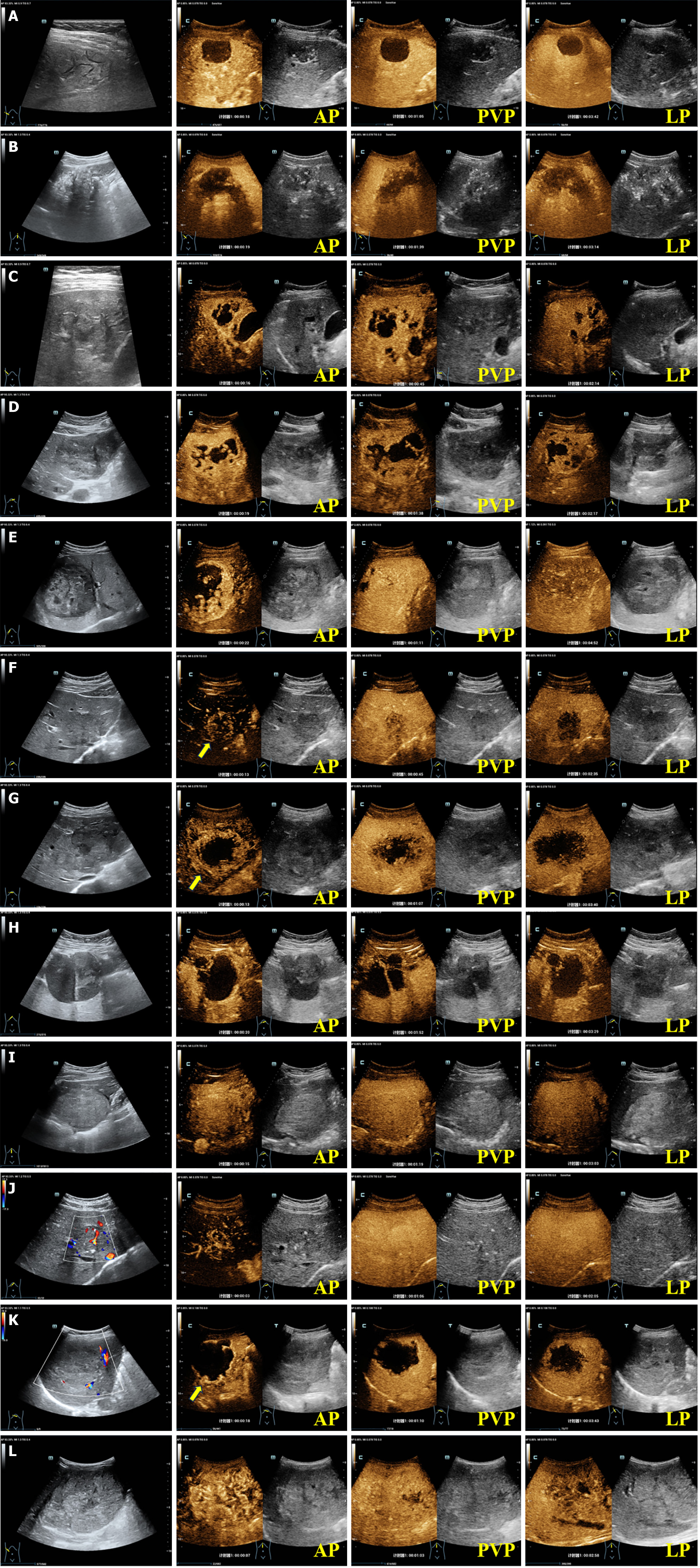
Figure 5 Contrast-enhanced ultrasound images of solid focal liver lesions.
A: A 37-year-old female patient presenting with a lesion of hepatic cystic echinococcosis 4, revealing no enhancement in the arterial phase (AP), portal venous phase (PVP), and late phase (LP); B: A 30-year-old female patient presenting with a hailstorm hepatic alveolar echinococcosis lesion, exhibiting irregular mild hyper-enhancement around the periphery in the AP and hypo-enhancement in the PVP and LP, resembling a “worm-eaten” appearance, with no significant enhancement observed in most of the internal regions; C: A 49-year-old female patient presenting with a lesion consistent with paragonimiasis, exhibiting heterogeneous enhancement in the AP, with non-enhancing reticular and “tunnel-like” areas internally; D: A 70-year-old female patient presenting with a liver abscess, showing hyper-enhancement of the cystic wall and internal septa in the AP, with multiple patches of non-enhancing liquefied necrotic areas, creating a “honeycomb-like” pattern; E: A 73-year-old male patient presenting with a lesion from hepatocellular carcinoma, exhibiting heterogeneous hyper-enhancement in the AP, iso-enhancement in the PVP, and hypo-enhancement in the LP; F: A 36-year-old male patient presenting with a lesion indicative of intrahepatic cholangiocarcinoma, showing irregular “rim-like” peripheral enhancement in the AP (the yellow arrow), washout in the PVP, and significant hypo-enhancement in the LP; G: A 40-year-old male patient presenting with liver metastasis, showing thick “rim-like” hyper-enhancement in the early AP (the yellow arrow), with washout in the early PVP and marked hypo-enhancement in the LP, presenting as a “bull's eye” sign; H: A 57-year-old female patient presenting with hepatic biliary cystadenoma. The lesion shows slight enhancement of the wall and septa in the AP, with some areas exhibiting thickened septa and dense nodular enhancement, followed by decreased enhancement in the PVP and LP; I: A 14-year-old female patient presenting with hepatocellular adenoma. The lesion shows slight hyper-enhancement in the AP and LP, and iso-enhancement in the LP; J: A 23-year-old male patient presenting with a lesion consistent with focal nodular hyperplasia, characterized by centrifugal enhancement in a “spoke-wheel” or “firework-like” pattern extending from the center to the periphery in the AP. The lesion shows slight hyper-enhancement in the PVP, and iso-enhancement in the LP; K: A 57-year-old female patient presenting with hepatic sclerosing hemangioma. The lesion shows discontinuous, nodular peripheral enhancement in the AP (the yellow arrow), with progressive partial or complete centripetal fill-in in the PVP and slight hyper-enhancement in the LP, as well as patchy non-enhancing areas observed internally; L: A 2-year-old male patient presenting with a lesion associated with hepatoblastoma, showing hyper-enhancement in the AP, slight hypo-enhancement in the PVP, and hypo-enhancement in the LP.
- Citation: Tao Y, Wang YF, Wang J, Long S, Seyler BC, Zhong XF, Lu Q. Pictorial review of hepatic echinococcosis: Ultrasound imaging and differential diagnosis. World J Gastroenterol 2024; 30(37): 4115-4131
- URL: https://www.wjgnet.com/1007-9327/full/v30/i37/4115.htm
- DOI: https://dx.doi.org/10.3748/wjg.v30.i37.4115


