Copyright
©The Author(s) 2024.
World J Gastroenterol. Aug 14, 2024; 30(30): 3574-3583
Published online Aug 14, 2024. doi: 10.3748/wjg.v30.i30.3574
Published online Aug 14, 2024. doi: 10.3748/wjg.v30.i30.3574
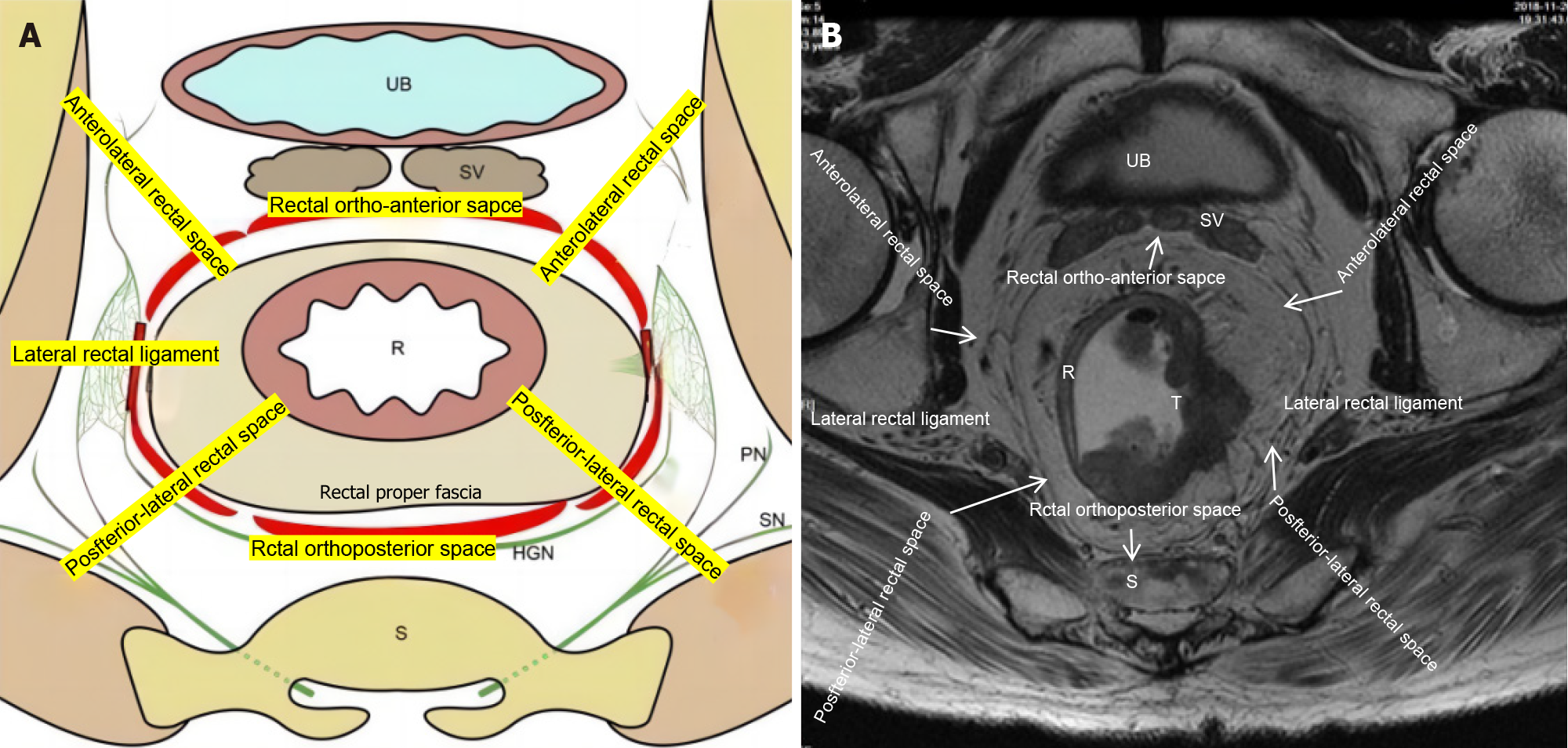
Figure 1 Schematic diagram of the "eight-zone method" of the pelvic floor fascia.
A: Schematic image of the “eight-zone method” of the pelvic floor fascia; B: Magnetic resonance image of the “eight-zone method” of the pelvic floor fascia. UB: Urinary bladder; SV: Seminal vesicle; R: Rectum; S: Sacrum; RN: Perineal nerve; SN: Sciatic nerve.
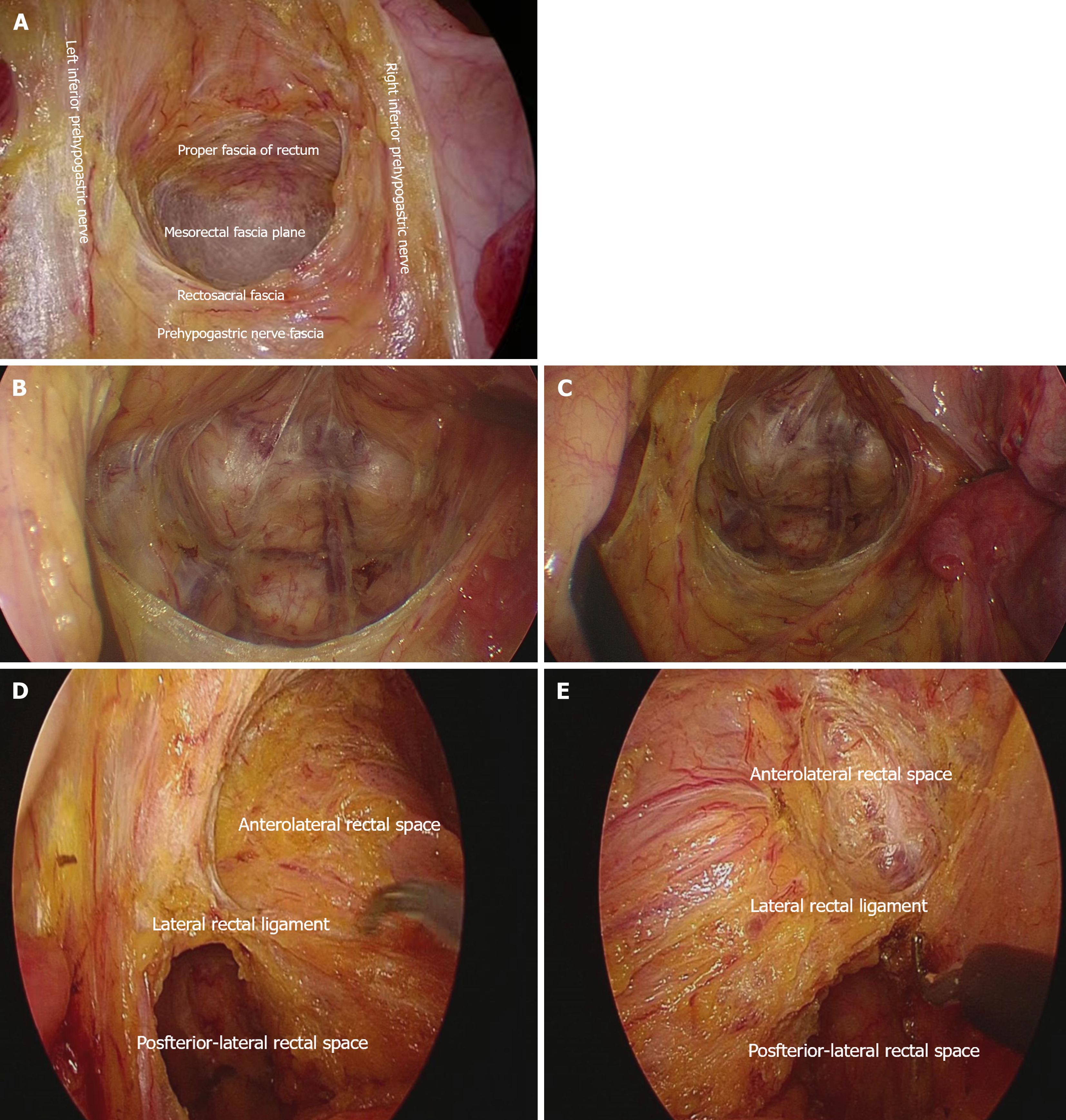
Figure 2 Posterior pelvic floor rectal, posterolateral and lateral rectal space.
A: Posterior pelvic floor rectal space; B: Left posterolateral space; C: Right posterolateral space; D: Left lateral rectal space; E: Right lateral rectal space.
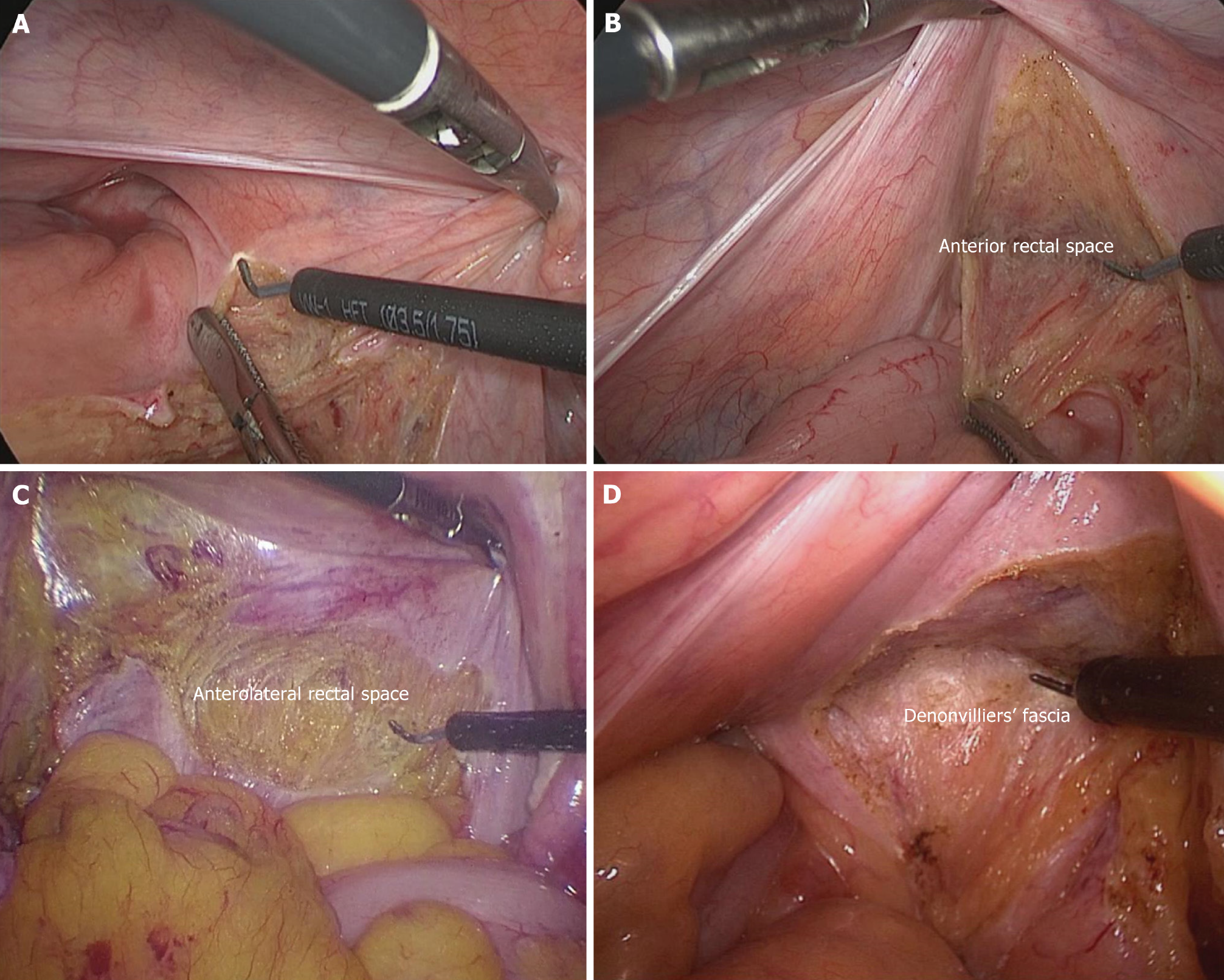
Figure 3 Anterior rectal space.
A: The pelvic floor peritoneum; B: Anterior rectal space; C: Anterolateral rectal space; D: Denonvilliers’ fascia.
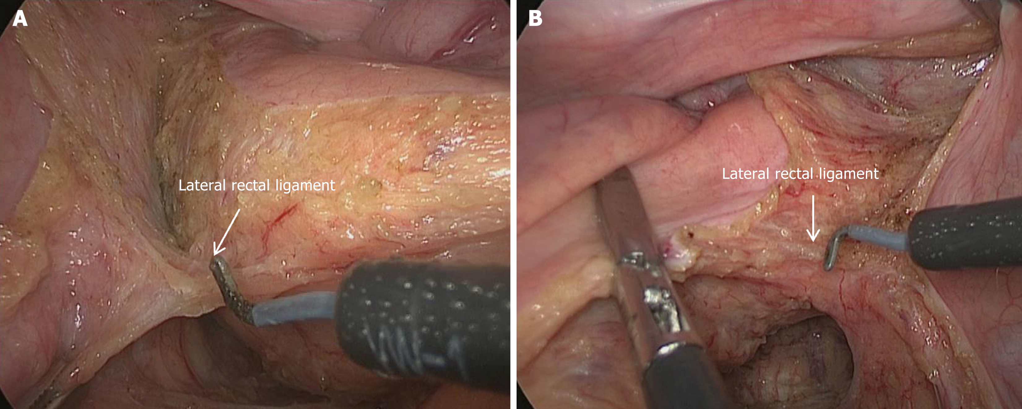
Figure 4 Separation of the left and right lateral rectal ligaments.
A: Left lateral rectal ligament; B: Right lateral rectal ligament.
- Citation: Chen C, Zhang X, Li X, Wang YL. Clinical application of eight-zone laparoscopic dissection strategy for rectal cancer: Experience and discussion. World J Gastroenterol 2024; 30(30): 3574-3583
- URL: https://www.wjgnet.com/1007-9327/full/v30/i30/3574.htm
- DOI: https://dx.doi.org/10.3748/wjg.v30.i30.3574


