Copyright
©The Author(s) 2024.
World J Gastroenterol. Jun 21, 2024; 30(23): 2981-2990
Published online Jun 21, 2024. doi: 10.3748/wjg.v30.i23.2981
Published online Jun 21, 2024. doi: 10.3748/wjg.v30.i23.2981
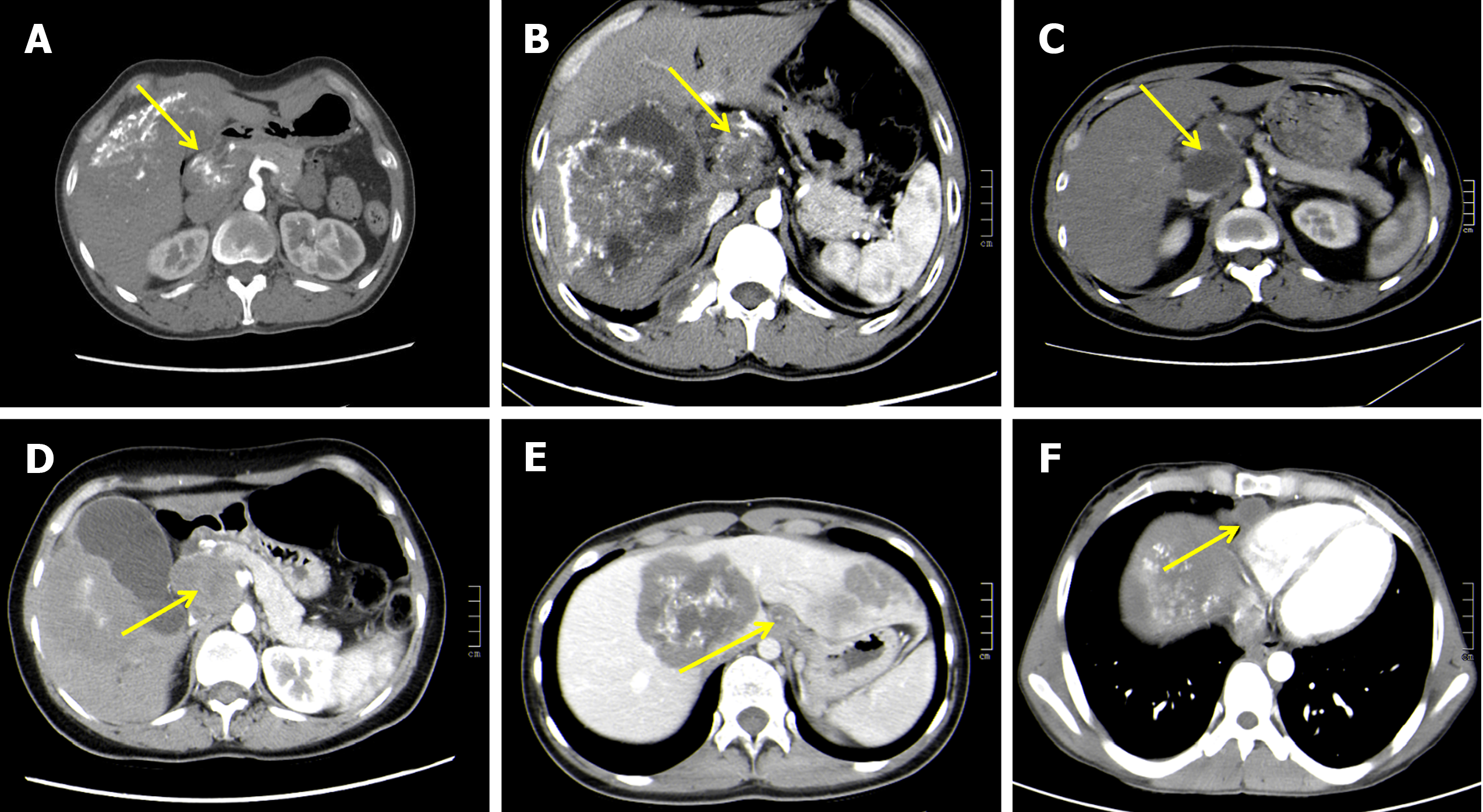
Figure 1 Computed tomography findings of lymph node metastasis in hepatic alveolar echinococcosis.
A: An enlarged mixed-density lymph node with multiple nodular, eggshell calcifications next to the portal vein in the first hepatic portal area and an unenhanced liquefied necrotic area in the lesion on an enhanced scan; B: A roundish mixed-density mass next to the common hepatic artery, with multiple sand-like, nodular calcifications within the lesion, with mild to moderate enhancement of the lesion on an enhanced scan and a hypoenhanced liquefied necrotic area within the lesion; C: Enlarged lymph nodes were observed at the bifurcation level of the splenic artery and common hepatic artery on the right side of the celiac trunk, with uniform density within them, and patchy but unenhanced areas of liquefied necrosis were seen within the lesion on enhancement scans; D: A mixed-density mass in the interstitial space of the abdominal aorta posterior to the pancreatic head with a speckled high-density shadow and a patchy slightly low-density shadow, heterogeneous enhancement on an enhanced scan, and no significant enhancement in the slightly low-density liquefied necrotic area within the lesion; E: An enlarged heterogeneously dense lymph node with nodular foci next to the abdominal aorta, with a mild to moderate enhanced lesion edge on an enhanced scan and no enhancement within the lesion; F: A roundish, slightly hypointense shadow in the right diaphragmatic angle with circular enhancement at the lesion edge on an enhanced scan and no significant.
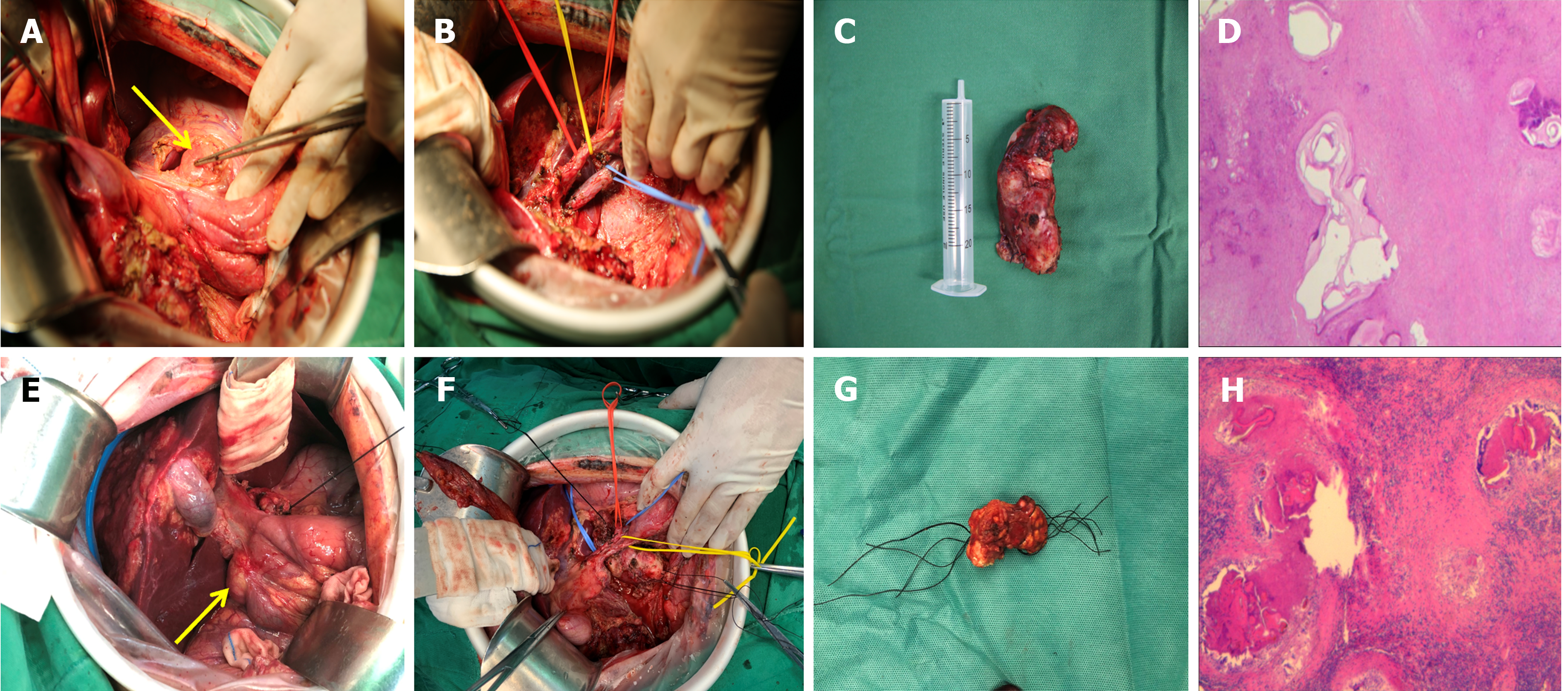
Figure 2 A 19-year-old male patient presented with intermittent epigastric pain and discomfort for 3 months and was diagnosed with hepatic alveolar echinococcosis combined with porta hepatis lymph node metastasis based on clinical examination and imaging studies.
A: Para-hepatoduodenal ligament lymph node enlargement was observed intraoperatively; B: Dissection of the hepatoduodenal ligament to skeletonization; C and D: Postoperative lymph node specimens and pathological sections; E-H: A 50-year-old female patient presented with right upper abdominal distension for over 4 years and was diagnosed with hepatic alveolar echinococcosis (AE) with posterior pancreatic head lymph node metastasis based on clinical examination and imaging findings. Posterior pancreatic head lymph node enlargement was observed intraoperatively (E). The regional lymph nodes were dissected to skeletonisation (F). Postoperative lymph node specimens and pathological sections (G and H).
- Citation: Aimaitijiang Y, Jiang TM, Shao YM, Aji T. Fifty-five cases of hepatic alveolar echinococcosis combined with lymph node metastasis: A retrospective study. World J Gastroenterol 2024; 30(23): 2981-2990
- URL: https://www.wjgnet.com/1007-9327/full/v30/i23/2981.htm
- DOI: https://dx.doi.org/10.3748/wjg.v30.i23.2981


