Copyright
©The Author(s) 2024.
World J Gastroenterol. May 7, 2024; 30(17): 2321-2331
Published online May 7, 2024. doi: 10.3748/wjg.v30.i17.2321
Published online May 7, 2024. doi: 10.3748/wjg.v30.i17.2321
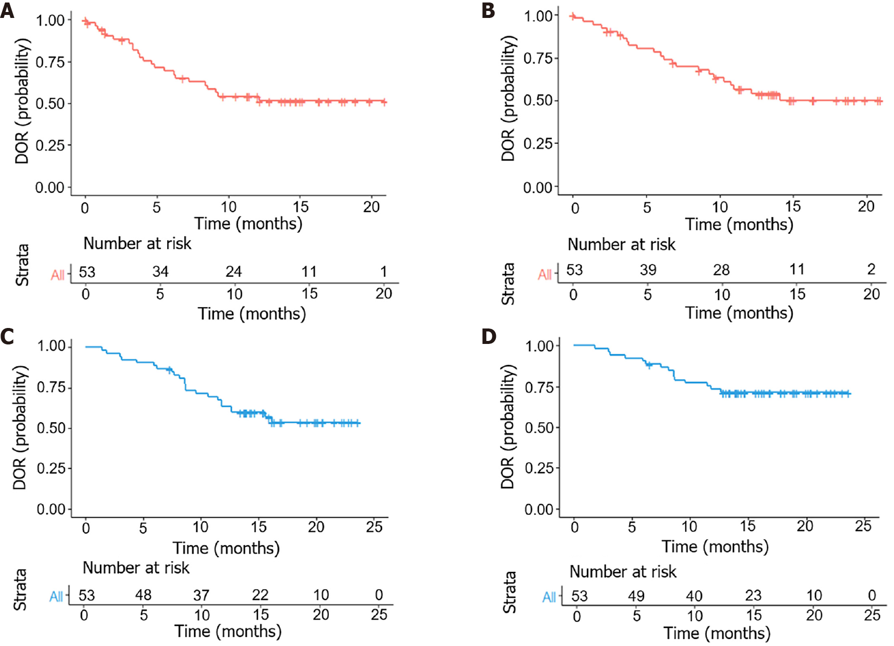
Figure 1 Prognostic outcomes of patients with hepatocellular carcinoma followed up within 25 months after treatment.
A: Median duration of response (DOR) using modified Response Evaluation Criteria in Solid Tumors; B: Median DOR using RECIST v1.1; C: Median progression-free survival; D: Median overall survival.
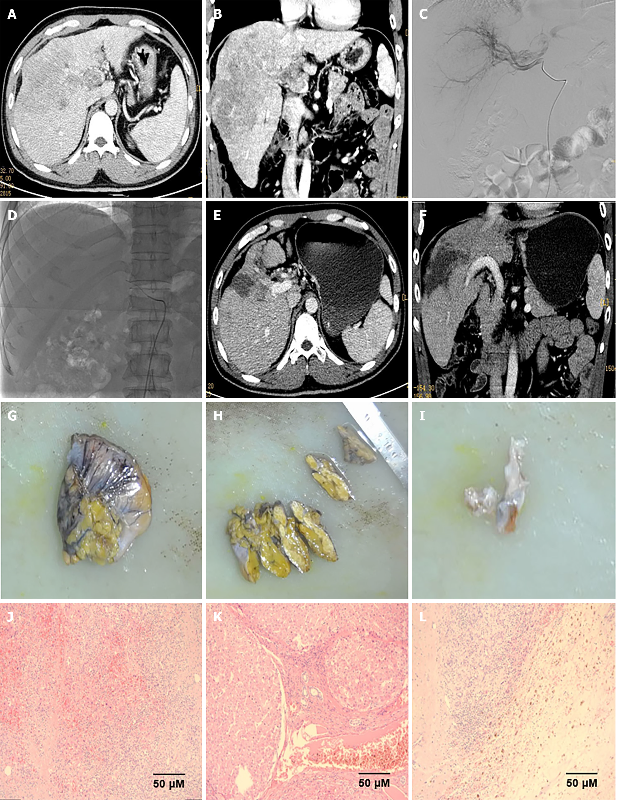
Figure 2 Imaging and pathological results of patients with hepatocellular carcinoma.
A and B: Massive hepatocellular carcinoma in the right lobe of the liver and tumor thrombus formation in the main portal vein and right branch; C: Digital subtraction angiography showing a mass in the right lobe of the liver; D: Microcatheter insertion into the appropriate hepatic artery for FOLFOX chemotherapy; E and F: After four rounds of hepatic arterial infusion chemotherapy, enhanced computed tomography of the upper abdomen revealed that the intrahepatic lesions were significantly reduced, the portal vein was reopened, and the portal vein tumor thrombus was significantly reduced; G-I: Gross pathological specimens after surgery; J-L: The tumor was completely necrotic with no residual cancer tissue, indicating complete pathological remission.
- Citation: Lin ZP, Hu XL, Chen D, Huang DB, Zou XG, Zhong H, Xu SX, Chen Y, Li XQ, Zhang J. Efficacy and safety of targeted therapy plus immunotherapy combined with hepatic artery infusion chemotherapy (FOLFOX) for unresectable hepatocarcinoma. World J Gastroenterol 2024; 30(17): 2321-2331
- URL: https://www.wjgnet.com/1007-9327/full/v30/i17/2321.htm
- DOI: https://dx.doi.org/10.3748/wjg.v30.i17.2321


